

chapter 8 nanomaterials: Synthesis and characterization
274
probe is then scanned over the surface of the material at distances
of a few nanometers above the surface (near field). Because the distance between the aperture and the sample is much smaller than
the diameter of the aperture, the illuminated area will be a function of the aperture dimensions and not limited by the diffraction
limit. In other words, NSOM resolution is limited by the size of the
aperture. In this way, by scanning a sample with the “near field” of a
focused light source, optical images with resolution around 50 nm
can be generated. Normally, fluorescent, topographical, and transmitted images are available (Figure 8.30). Currently, NSOM is used
in a wide range of fields to observe nanoscale features. In addition,
because NSOM images the surface point by point, a topographical
image of the surface can be resolved at the same time as the optical
image.
One other important light microscopy technique is the confocal
scanning light microscope. Invented by Marvin Minsky in the 1950s,
confocal scanning microscopy is based on the principle that the
existence of a pinhole in front of a detector only allows the traveling
signal from the focal plane of the objective to enter the detector.
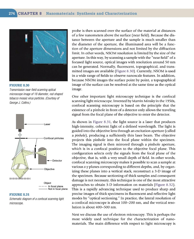
As shown in Figure 8.31, the light source is a laser that produces
high-intensity, coherent light of a defined wavelength. The light is
guided into the objective lens through an excitation aperture (called
a pinhole), producing a sufficiently thin laser beam. The objective
projects this pinhole into the focal plane within the specimen.
The imaging signal is then mirrored through a pinhole aperture,
which is in a confocal position to the objective focal plane. This
configuration selects only the signals from the focal plane of the
objective, that is, with a very small depth of field. In other words,
confocal scanning microscopy makes it possible to scan a sample at
various x-y planes corresponding to different depths, and, by organizing these planes into a vertical stack, reconstruct a 3-D image of
the specimen. Because sectioning of thick samples and consequent
damage is not necessary, this technique is one of the most attractive
approaches to obtain 3-D information on materials (Figure 8.32).
This is a rapidly advancing technique used to produce sharp and
precise images of thick specimens in fluorescent and reflective light
modes by “optical sectioning.” In practice, the lateral resolution of
a confocal microscope is about 100−200 nm, and the vertical resolution is about 400−500 nm.
Next we discuss the use of electron microscopy. This is perhaps the
most widely used technique for the characterization of nanomaterials. The main difference with respect to light microscopy is
Figure 8.30
Transmission near-field scanning optical
microscope image of 18 diameter, rod-shaped
tobacco mosaic virus particles. (Courtesy of
George J. Collins.)
Figure 8.31
Schematic diagram of a confocal scanning light
microscope.
Confocal pinholes
Laser
Objective
Detector
Object
In focal plane
Not in focal plane
274
probe is then scanned over the surface of the material at distances
of a few nanometers above the surface (near field). Because the distance between the aperture and the sample is much smaller than
the diameter of the aperture, the illuminated area will be a function of the aperture dimensions and not limited by the diffraction
limit. In other words, NSOM resolution is limited by the size of the
aperture. In this way, by scanning a sample with the “near field” of a
focused light source, optical images with resolution around 50 nm
can be generated. Normally, fluorescent, topographical, and transmitted images are available (Figure 8.30). Currently, NSOM is used
in a wide range of fields to observe nanoscale features. In addition,
because NSOM images the surface point by point, a topographical
image of the surface can be resolved at the same time as the optical
image.
One other important light microscopy technique is the confocal
scanning light microscope. Invented by Marvin Minsky in the 1950s,
confocal scanning microscopy is based on the principle that the
existence of a pinhole in front of a detector only allows the traveling
signal from the focal plane of the objective to enter the detector.
As shown in Figure 8.31, the light source is a laser that produces
high-intensity, coherent light of a defined wavelength. The light is
guided into the objective lens through an excitation aperture (called
a pinhole), producing a sufficiently thin laser beam. The objective
projects this pinhole into the focal plane within the specimen.
The imaging signal is then mirrored through a pinhole aperture,
which is in a confocal position to the objective focal plane. This
configuration selects only the signals from the focal plane of the
objective, that is, with a very small depth of field. In other words,
confocal scanning microscopy makes it possible to scan a sample at
various x-y planes corresponding to different depths, and, by organizing these planes into a vertical stack, reconstruct a 3-D image of
the specimen. Because sectioning of thick samples and consequent
damage is not necessary, this technique is one of the most attractive
approaches to obtain 3-D information on materials (Figure 8.32).
This is a rapidly advancing technique used to produce sharp and
precise images of thick specimens in fluorescent and reflective light
modes by “optical sectioning.” In practice, the lateral resolution of
a confocal microscope is about 100−200 nm, and the vertical resolution is about 400−500 nm.
Next we discuss the use of electron microscopy. This is perhaps the
most widely used technique for the characterization of nanomaterials. The main difference with respect to light microscopy is
Figure 8.30
Transmission near-field scanning optical
microscope image of 18 diameter, rod-shaped
tobacco mosaic virus particles. (Courtesy of
George J. Collins.)
Figure 8.31
Schematic diagram of a confocal scanning light
microscope.
Confocal pinholes
Laser
Objective
Detector
Object
In focal plane
Not in focal plane