

273
type (0-D, 1-D, 2-D, and 3-D), one or more techniques can be used
to extract the desirable information. A simple way of categorizing
the characterization methods is to consider imaging and analytical techniques. Imaging involves some kind of microscopy, whereas
analysis involves some type of spectroscopy.
Let’s begin with the imaging techniques. These methods can involve
light, electrons, ions, or scanning probes. With respect to light microscopes, the most common approach for imaging nanomaterials is
the so-called near-field scanning optical microscope (NSOM; see Figure
8.29). In this type of microscopy a subwavelength aperture (smaller
than the wavelength of light) is placed in close proximity to the
sample (∼10 nm). In addition, a light source is used as a scanning
probe. To achieve good resolution, the probe is highly focused. The
Characterization of Nanomaterials
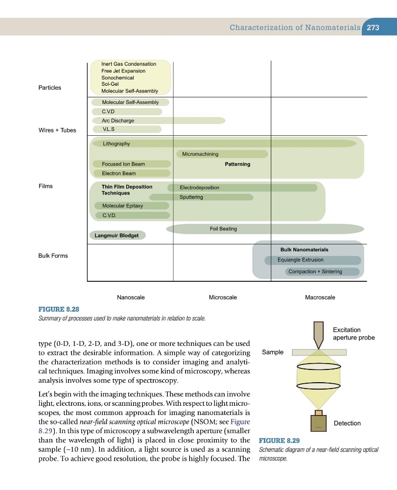
Figure 8.28
Summary of processes used to make nanomaterials in relation to scale.
Particles
Wires + Tubes
Films
Bulk Forms
Nanoscale
Microscale
Macroscale
Inert Gas Condensation
Molecular Self-Assembly
Sonochemical
Sol-Gel
C.V.D
Arc Discharge
V.L.S
Patterning
Lithography
Micromachining
Thin Film Deposition
Techniques
Electrodeposition
Sputtering
Molecular Epitaxy
Langmuir Blodget
Equiangle Extrusion
Compaction + Sintering
Bulk Nanomaterials
Free Jet Expansion
Molecular Self-Assembly
Focused Ion Beam
Electron Beam
C.V.D.
Foil Beating
Sample
Detection
Excitation
aperture probe
Figure 8.29
Schematic diagram of a near-field scanning optical
microscope.
type (0-D, 1-D, 2-D, and 3-D), one or more techniques can be used
to extract the desirable information. A simple way of categorizing
the characterization methods is to consider imaging and analytical techniques. Imaging involves some kind of microscopy, whereas
analysis involves some type of spectroscopy.
Let’s begin with the imaging techniques. These methods can involve
light, electrons, ions, or scanning probes. With respect to light microscopes, the most common approach for imaging nanomaterials is
the so-called near-field scanning optical microscope (NSOM; see Figure
8.29). In this type of microscopy a subwavelength aperture (smaller
than the wavelength of light) is placed in close proximity to the
sample (∼10 nm). In addition, a light source is used as a scanning
probe. To achieve good resolution, the probe is highly focused. The
Characterization of Nanomaterials
Figure 8.28
Summary of processes used to make nanomaterials in relation to scale.
Particles
Wires + Tubes
Films
Bulk Forms
Nanoscale
Microscale
Macroscale
Inert Gas Condensation
Molecular Self-Assembly
Sonochemical
Sol-Gel
C.V.D
Arc Discharge
V.L.S
Patterning
Lithography
Micromachining
Thin Film Deposition
Techniques
Electrodeposition
Sputtering
Molecular Epitaxy
Langmuir Blodget
Equiangle Extrusion
Compaction + Sintering
Bulk Nanomaterials
Free Jet Expansion
Molecular Self-Assembly
Focused Ion Beam
Electron Beam
C.V.D.
Foil Beating
Sample
Detection
Excitation
aperture probe
Figure 8.29
Schematic diagram of a near-field scanning optical
microscope.