

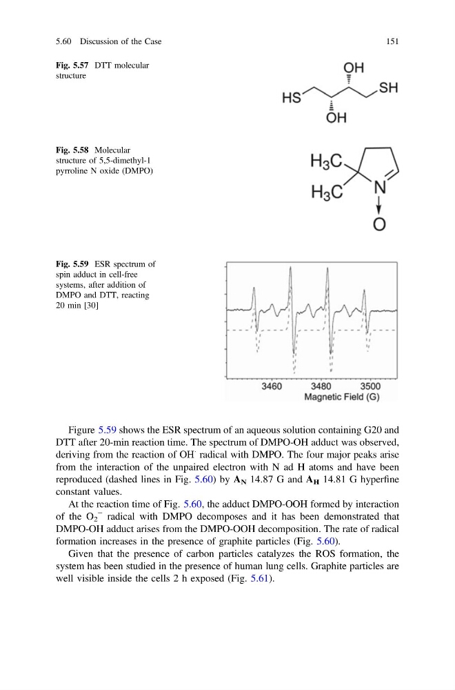
Figure 5.59 shows the ESR spectrum of an aqueous solution containing G20 and
DTT after 20-min reaction time. The spectrum of DMPO-OH adduct was observed,
deriving from the reaction of OH
Á radical with DMPO. The four major peaks arise
from the interaction of the unpaired electron with N ad H atoms and have been
reproduced (dashed lines in Fig. 5.60) by A N 14.87 G and A H 14.81 G hyperfine
constant values.
At the reaction time of Fig. 5.60, the adduct DMPO-OOH formed by interaction
of the O 2
− radical with DMPO decomposes and it has been demonstrated that
DMPO-OH adduct arises from the DMPO-OOH decomposition. The rate of radical
formation increases in the presence of graphite particles (Fig. 5.60).
Given that the presence of carbon particles catalyzes the ROS formation, the
system has been studied in the presence of human lung cells. Graphite particles are
well visible inside the cells 2 h exposed (Fig. 5.61).
Fig. 5.57 DTT molecular
structure
Fig. 5.58 Molecular
structure of 5,5-dimethyl-1
pyrroline N oxide (DMPO)
Fig. 5.59 ESR spectrum of
spin adduct in cell-free
systems, after addition of
DMPO and DTT, reacting
20 min [30]
5.60 Discussion of the Case
151
DTT after 20-min reaction time. The spectrum of DMPO-OH adduct was observed,
deriving from the reaction of OH
Á radical with DMPO. The four major peaks arise
from the interaction of the unpaired electron with N ad H atoms and have been
reproduced (dashed lines in Fig. 5.60) by A N 14.87 G and A H 14.81 G hyperfine
constant values.
At the reaction time of Fig. 5.60, the adduct DMPO-OOH formed by interaction
of the O 2
− radical with DMPO decomposes and it has been demonstrated that
DMPO-OH adduct arises from the DMPO-OOH decomposition. The rate of radical
formation increases in the presence of graphite particles (Fig. 5.60).
Given that the presence of carbon particles catalyzes the ROS formation, the
system has been studied in the presence of human lung cells. Graphite particles are
well visible inside the cells 2 h exposed (Fig. 5.61).
Fig. 5.57 DTT molecular
structure
Fig. 5.58 Molecular
structure of 5,5-dimethyl-1
pyrroline N oxide (DMPO)
Fig. 5.59 ESR spectrum of
spin adduct in cell-free
systems, after addition of
DMPO and DTT, reacting
20 min [30]
5.60 Discussion of the Case
151