

3.3. MrCAOSCOPY
57
and the latter monitors the force exerted between Ihc surfacc and the probe tip. The
AFM, like the STM, has two modes of operation. The AFM can operate in a close
contact mode in which the core-to-core repulsive forces with the surface dominate,
or in a greater separation %oncontact" mude in which the relevant force is the
gradient of the van der Waals potential. As in the STM casc, a piczoelectric Scanner
is used. The vertical motions of the tip during the scanning may he monitored by
the interfemce pattern of a light beam from an optical fiber, as shown in the upper
diagmam of the figure, or by the reflection of a laser heam, ns shown in the enlarged
view of the probe tip in the lower diagram of the figure. The ntomic force microscope
is sensitive to the vertical component of the surface forces. A related hut more
versatile device called I fricfiorifun-e microscope, also somctirncs rcfcrred to as a
JaremlJk-ce mictmcope, simultaneously measures kith normal and lateral forces of
thc surface on the tip.
At1 three of these scanning microscopes can provide information on the
topography and defect structure of a surface over distances close t~ the atomic
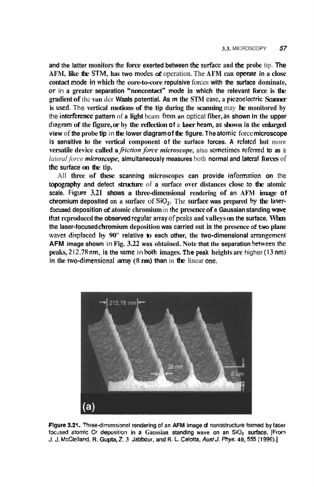
scale. Figure 3.21 shows a three-dimensional rendering of an AFM image of
chromium deposited on a surface of SiO,. The surface was prepared hy the l a w -
focused deposition of atomic chromium in the presence ofa Gaussian standing wave
that rcproduccd the observed regular array ofpeaks and valleys on the surface. When
the laser-focused chromium deposition was carried out in the presence of two plane
wwes displaced by 90" relative to each other, the two-dimensional amngemcnt
AFM image shown in Fig. 3.22 was obtained. Note that the separation heween thc
peaks, 212.78 nm, is the same in hoth images. The peak heights are higher (I3 nm)
in the two-dimensionaI array (R nm) than in the linear QIE.
Flgure 3.21. Threedimenslonal rendering of an AFM image of nanostructure formed by raset
focused atomic Q deposittan in a Gaussian standing wave on an SiQz surface. [Frm
J. J. Mcclelland, R. Gupta, Z. 3 Jabbour. and R. L. Celotta. Awsf J. Phgrs. 49, 555 (199Q.1
57
and the latter monitors the force exerted between Ihc surfacc and the probe tip. The
AFM, like the STM, has two modes of operation. The AFM can operate in a close
contact mode in which the core-to-core repulsive forces with the surface dominate,
or in a greater separation %oncontact" mude in which the relevant force is the
gradient of the van der Waals potential. As in the STM casc, a piczoelectric Scanner
is used. The vertical motions of the tip during the scanning may he monitored by
the interfemce pattern of a light beam from an optical fiber, as shown in the upper
diagmam of the figure, or by the reflection of a laser heam, ns shown in the enlarged
view of the probe tip in the lower diagram of the figure. The ntomic force microscope
is sensitive to the vertical component of the surface forces. A related hut more
versatile device called I fricfiorifun-e microscope, also somctirncs rcfcrred to as a
JaremlJk-ce mictmcope, simultaneously measures kith normal and lateral forces of
thc surface on the tip.
At1 three of these scanning microscopes can provide information on the
topography and defect structure of a surface over distances close t~ the atomic
scale. Figure 3.21 shows a three-dimensional rendering of an AFM image of
chromium deposited on a surface of SiO,. The surface was prepared hy the l a w -
focused deposition of atomic chromium in the presence ofa Gaussian standing wave
that rcproduccd the observed regular array ofpeaks and valleys on the surface. When
the laser-focused chromium deposition was carried out in the presence of two plane
wwes displaced by 90" relative to each other, the two-dimensional amngemcnt
AFM image shown in Fig. 3.22 was obtained. Note that the separation heween thc
peaks, 212.78 nm, is the same in hoth images. The peak heights are higher (I3 nm)
in the two-dimensionaI array (R nm) than in the linear QIE.
Flgure 3.21. Threedimenslonal rendering of an AFM image of nanostructure formed by raset
focused atomic Q deposittan in a Gaussian standing wave on an SiQz surface. [Frm
J. J. Mcclelland, R. Gupta, Z. 3 Jabbour. and R. L. Celotta. Awsf J. Phgrs. 49, 555 (199Q.1