

8.3. LUMINESCENCE
21 7
Excitatior I [eV]: 2.175
2.214
2.255
2.296
2.318
2.340
2.385
2.431
- &
2.455
2.480
2.531
~ I I I I I I I I I I I I I I
Photon Energy (eV)
1.4 1.6 1.8 2 2.2 2.4 2.6 2.8
(a)
Excitation [ev]:
2.175
2.431
& 2.455
1.8 2 2.2 2.4 2.6
Photon Energy (eV)
(b)
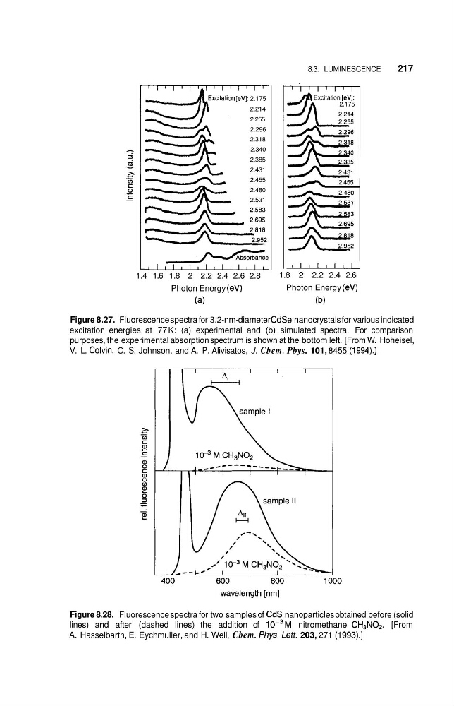
Figure 8.27. Fluorescence spectra for 3.2-nm-diameter CdSe nanocrystals for various indicated
excitation energies at 77 K: (a) experimental and (b) simulated spectra. For comparison
purposes, the experimental absorption spectrum is shown at the bottom left. [From W. Hoheisel,
V. L. Colvin, C. S. Johnson, and A. P. Alivisatos, J. Cbem. Pbys. 101, 8455 (1994).]
Figure 8.28. Fluorescence spectra for two samples of CdS nanoparticles obtained before (solid
lines) and after (dashed lines) the addition of 10-3M nitromethane CH3N02. [From
A. Hasselbarth, E. Eychmuller, and H. Well, Cbem. Pbys. Lett. 203, 271 (1993).]
21 7
Excitatior I [eV]: 2.175
2.214
2.255
2.296
2.318
2.340
2.385
2.431
- &
2.455
2.480
2.531
~ I I I I I I I I I I I I I I
Photon Energy (eV)
1.4 1.6 1.8 2 2.2 2.4 2.6 2.8
(a)
Excitation [ev]:
2.175
2.431
& 2.455
1.8 2 2.2 2.4 2.6
Photon Energy (eV)
(b)
Figure 8.27. Fluorescence spectra for 3.2-nm-diameter CdSe nanocrystals for various indicated
excitation energies at 77 K: (a) experimental and (b) simulated spectra. For comparison
purposes, the experimental absorption spectrum is shown at the bottom left. [From W. Hoheisel,
V. L. Colvin, C. S. Johnson, and A. P. Alivisatos, J. Cbem. Pbys. 101, 8455 (1994).]
Figure 8.28. Fluorescence spectra for two samples of CdS nanoparticles obtained before (solid
lines) and after (dashed lines) the addition of 10-3M nitromethane CH3N02. [From
A. Hasselbarth, E. Eychmuller, and H. Well, Cbem. Pbys. Lett. 203, 271 (1993).]