

12
1 Redox Proteins and Bioelectrocatalysis
1.2.5 Ion-Sulfur Clusters [1, 7]
Non-heme iron proteins contain ion-sulfur (FeS) clusters, which are also the
common type of single-electron carrier. Cys, His, Glu, Asp, and Tyr are the most
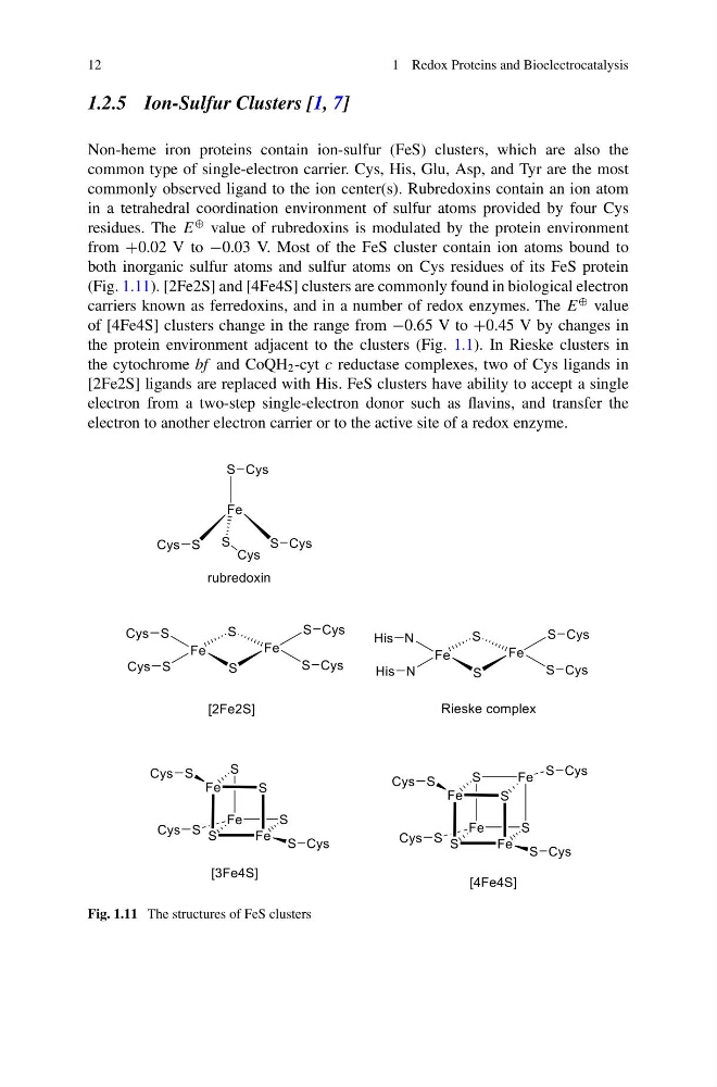
commonly observed ligand to the ion center(s). Rubredoxins contain an ion atom
in a tetrahedral coordination environment of sulfur atoms provided by four Cys
residues. The E
⊕ value of rubredoxins is modulated by the protein environment
from +0.02 V to −0.03 V. Most of the FeS cluster contain ion atoms bound to
both inorganic sulfur atoms and sulfur atoms on Cys residues of its FeS protein
(Fig. 1.11). [2Fe2S] and [4Fe4S] clusters are commonly found in biological electron
carriers known as ferredoxins, and in a number of redox enzymes. The E
⊕ value
of [4Fe4S] clusters change in the range from −0.65 V to +0.45 V by changes in
the protein environment adjacent to the clusters (Fig. 1.1). In Rieske clusters in
the cytochrome bf and CoQH 2 -cyt c reductase complexes, two of Cys ligands in
[2Fe2S] ligands are replaced with His. FeS clusters have ability to accept a single
electron from a two-step single-electron donor such as flavins, and transfer the
electron to another electron carrier or to the active site of a redox enzyme.
Fig. 1.11 The structures of FeS clusters
1 Redox Proteins and Bioelectrocatalysis
1.2.5 Ion-Sulfur Clusters [1, 7]
Non-heme iron proteins contain ion-sulfur (FeS) clusters, which are also the
common type of single-electron carrier. Cys, His, Glu, Asp, and Tyr are the most
commonly observed ligand to the ion center(s). Rubredoxins contain an ion atom
in a tetrahedral coordination environment of sulfur atoms provided by four Cys
residues. The E
⊕ value of rubredoxins is modulated by the protein environment
from +0.02 V to −0.03 V. Most of the FeS cluster contain ion atoms bound to
both inorganic sulfur atoms and sulfur atoms on Cys residues of its FeS protein
(Fig. 1.11). [2Fe2S] and [4Fe4S] clusters are commonly found in biological electron
carriers known as ferredoxins, and in a number of redox enzymes. The E
⊕ value
of [4Fe4S] clusters change in the range from −0.65 V to +0.45 V by changes in
the protein environment adjacent to the clusters (Fig. 1.1). In Rieske clusters in
the cytochrome bf and CoQH 2 -cyt c reductase complexes, two of Cys ligands in
[2Fe2S] ligands are replaced with His. FeS clusters have ability to accept a single
electron from a two-step single-electron donor such as flavins, and transfer the
electron to another electron carrier or to the active site of a redox enzyme.
Fig. 1.11 The structures of FeS clusters