

1.2 Redox Components
11
single Fe ion coordinated in the center. There are three different types, a, b, and c,
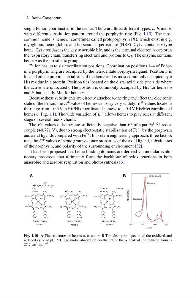
with different substitution pattern around the porphyrin ring (Fig. 1.10). The most
common heme is heme b (sometimes called protoporphyrin IX), which exist in e.g.
myoglobin, hemoglobin, and horseradish peroxidase (HRP). Cyt c contains c-type
heme. Cyt c oxidase is the key to aerobic life, and is the terminal electron acceptor in
the respiratory chain, transferring electrons and protons to O 2 . The enzyme contains
heme a as the prosthetic group.
Fe ion has up to six coordination positions. Coordination positions 1–4 of Fe ion
in a porphyrin ring are occupied by the tetradentate porphyrin ligand. Position 5 is
located on the proximal axial side of the heme and is most commonly occupied by a
His residue in a protein. Position 6 is located on the distal axial side (the side where
the active site is located). The position is commonly occupied by His for hemes a
and b, but usually Met for heme c.
Because these substituents are directly attached to the ring and affect the electronic
state of the Fe ion, the E
⊕ value of hemes can vary very widely; E
⊕ values locate in
the range from −0.3 V in His/His coordinated hemes c to +0.4 V His/Met coordinated
hemes c (Fig. 1.1). The wide variation of E
⊕ allows hemes to play roles at different
stage of several redox chains.
The E
⊕ values of hemes are sufficiently negative than E
◦ of aqua Fe
3+/2+ redox
couple (+0.771 V), due to strong electrostatic stabilization of Fe
3+ by the porphyrin
and axial ligands compared with Fe
2+ . In protein engineering approach, three factors
tune the E
⊕ values of heme groups: donor properties of the axial ligand, substituents
of the porphyrin, and polarity of the surrounding environment [32].
It has been proposed that heme-binding domains are derived via modular evolutionary processes that ultimately form the backbone of redox reactions in both
anaerobic and aerobic respiration and photosynthesis [31].
A
B
Fig. 1.10 A The structures of hemes a, b, and c. B The absorption spectra of the oxidized and
reduced cyt c at pH 7.0. The molar absorption coefficient of the α peak of the reduced form is
27.7 cm 2 mol −1
11
single Fe ion coordinated in the center. There are three different types, a, b, and c,
with different substitution pattern around the porphyrin ring (Fig. 1.10). The most
common heme is heme b (sometimes called protoporphyrin IX), which exist in e.g.
myoglobin, hemoglobin, and horseradish peroxidase (HRP). Cyt c contains c-type
heme. Cyt c oxidase is the key to aerobic life, and is the terminal electron acceptor in
the respiratory chain, transferring electrons and protons to O 2 . The enzyme contains
heme a as the prosthetic group.
Fe ion has up to six coordination positions. Coordination positions 1–4 of Fe ion
in a porphyrin ring are occupied by the tetradentate porphyrin ligand. Position 5 is
located on the proximal axial side of the heme and is most commonly occupied by a
His residue in a protein. Position 6 is located on the distal axial side (the side where
the active site is located). The position is commonly occupied by His for hemes a
and b, but usually Met for heme c.
Because these substituents are directly attached to the ring and affect the electronic
state of the Fe ion, the E
⊕ value of hemes can vary very widely; E
⊕ values locate in
the range from −0.3 V in His/His coordinated hemes c to +0.4 V His/Met coordinated
hemes c (Fig. 1.1). The wide variation of E
⊕ allows hemes to play roles at different
stage of several redox chains.
The E
⊕ values of hemes are sufficiently negative than E
◦ of aqua Fe
3+/2+ redox
couple (+0.771 V), due to strong electrostatic stabilization of Fe
3+ by the porphyrin
and axial ligands compared with Fe
2+ . In protein engineering approach, three factors
tune the E
⊕ values of heme groups: donor properties of the axial ligand, substituents
of the porphyrin, and polarity of the surrounding environment [32].
It has been proposed that heme-binding domains are derived via modular evolutionary processes that ultimately form the backbone of redox reactions in both
anaerobic and aerobic respiration and photosynthesis [31].
A
B
Fig. 1.10 A The structures of hemes a, b, and c. B The absorption spectra of the oxidized and
reduced cyt c at pH 7.0. The molar absorption coefficient of the α peak of the reduced form is
27.7 cm 2 mol −1