

1.2 Redox Components
13
1.2.6 Copper [33]
Copper proteins and enzymes contain redox active copper ion(s) in their active sites
in a permanently coordinated state. The functions of copper proteins are restricted
to electron transfer port in photosynthesis and nitrogen metabolism and in oxygen
transport, suggesting that copper ion gained biological importance only after the
oxygenation in the atmosphere. Cu
+ is a soft acid in the Person’s HSAB (hard and soft
acid base) concept and prefers Cys, and Met. The harder acid Cu
2+ can be coordinated
by Tyr, Ser, Thr, His, OH
− and H 2 O. In addition, when no steric hindrance occurs, Cu
+
prefers tetrahedral coordination, while the ligand of Cu
2+ arrange in a square planar
configuration. In the copper ion binding sites of copper proteins, both their ligands
and conformations considerably deviate from those preferred by Cu
2+ , leading to a
destabilization of the oxidized state and then a rise in the E
⊕ value of copper proteins
and to reduction of the reorganization energy associated with electron transfer. There
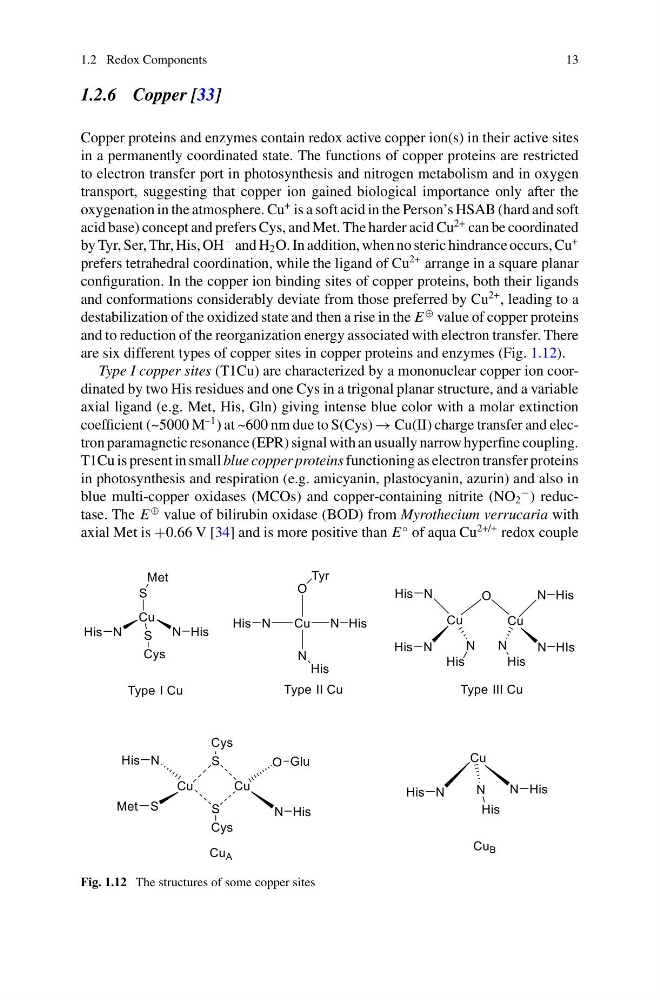
are six different types of copper sites in copper proteins and enzymes (Fig. 1.12).
Type I copper sites (T1Cu) are characterized by a mononuclear copper ion coordinated by two His residues and one Cys in a trigonal planar structure, and a variable
axial ligand (e.g. Met, His, Gln) giving intense blue color with a molar extinction
coefficient (~5000 M
–1 ) at ~600 nm due to S(Cys) → Cu(II) charge transfer and electron paramagnetic resonance (EPR) signal with an usually narrow hyperfine coupling.
T1Cu is present in small blue copper proteins functioning as electron transfer proteins
in photosynthesis and respiration (e.g. amicyanin, plastocyanin, azurin) and also in
blue multi-copper oxidases (MCOs) and copper-containing nitrite (NO 2
− ) reductase. The E
⊕ value of bilirubin oxidase (BOD) from Myrothecium verrucaria with
axial Met is +0.66 V [34] and is more positive than E
◦ of aqua Cu
2+/+ redox couple
Fig. 1.12 The structures of some copper sites
13
1.2.6 Copper [33]
Copper proteins and enzymes contain redox active copper ion(s) in their active sites
in a permanently coordinated state. The functions of copper proteins are restricted
to electron transfer port in photosynthesis and nitrogen metabolism and in oxygen
transport, suggesting that copper ion gained biological importance only after the
oxygenation in the atmosphere. Cu
+ is a soft acid in the Person’s HSAB (hard and soft
acid base) concept and prefers Cys, and Met. The harder acid Cu
2+ can be coordinated
by Tyr, Ser, Thr, His, OH
− and H 2 O. In addition, when no steric hindrance occurs, Cu
+
prefers tetrahedral coordination, while the ligand of Cu
2+ arrange in a square planar
configuration. In the copper ion binding sites of copper proteins, both their ligands
and conformations considerably deviate from those preferred by Cu
2+ , leading to a
destabilization of the oxidized state and then a rise in the E
⊕ value of copper proteins
and to reduction of the reorganization energy associated with electron transfer. There
are six different types of copper sites in copper proteins and enzymes (Fig. 1.12).
Type I copper sites (T1Cu) are characterized by a mononuclear copper ion coordinated by two His residues and one Cys in a trigonal planar structure, and a variable
axial ligand (e.g. Met, His, Gln) giving intense blue color with a molar extinction
coefficient (~5000 M
–1 ) at ~600 nm due to S(Cys) → Cu(II) charge transfer and electron paramagnetic resonance (EPR) signal with an usually narrow hyperfine coupling.
T1Cu is present in small blue copper proteins functioning as electron transfer proteins
in photosynthesis and respiration (e.g. amicyanin, plastocyanin, azurin) and also in
blue multi-copper oxidases (MCOs) and copper-containing nitrite (NO 2
− ) reductase. The E
⊕ value of bilirubin oxidase (BOD) from Myrothecium verrucaria with
axial Met is +0.66 V [34] and is more positive than E
◦ of aqua Cu
2+/+ redox couple
Fig. 1.12 The structures of some copper sites