

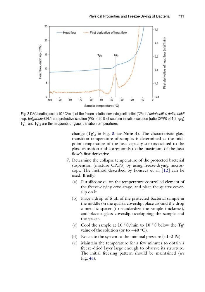
change (Tg
0
2 in Fig. 3, see Note 4). The characteristic glass
transition temperature of samples is determined as the midpoint temperature of the heat capacity step associated to the
glass transition and corresponds to the maximum of the heat
flow’s first derivative.
7. Determine the collapse temperature of the protected bacterial
suspension (mixture CP:PS) by using freeze-drying microscopy. The method described by Fonseca et al. [12] can be
used. Briefly:
(a) Put silicone oil on the temperature-controlled element of
the freeze-drying cryo-stage, and place the quartz coverslip on it.
(b) Place a drop of 5 μL of the protected bacterial sample in
the middle on the quartz coverslip, place around the drop
a metallic spacer (to standardize the sample thickness),
and place a glass coverslip overlapping the sample and
the spacer.
(c) Cool the sample at 10
C/min to 10
C below the Tg
0
value of the solution (or to À40
C).
(d) Evacuate the system to the minimal pressure (~1–2 Pa).
(e) Maintain the temperature for a few minutes to obtain a
freeze-dried layer large enough to observe its structure.
The initial freezing pattern should be maintained (see
Fig. 4a).
Fig. 3 DSC heating scan (10
C/min) of the frozen solution involving cell pellet (CP) of Lactobacillus delbrueckii
ssp. bulgaricus CFL1 and protective solution (PS) of 20% of sucrose in saline solution (ratio CP/PS of 1:2, g/g)
Tg
0
1 and Tg
0
2 are the midpoints of glass transition temperatures
Physical Properties and Freeze-Drying of Bacteria
711
0
2 in Fig. 3, see Note 4). The characteristic glass
transition temperature of samples is determined as the midpoint temperature of the heat capacity step associated to the
glass transition and corresponds to the maximum of the heat
flow’s first derivative.
7. Determine the collapse temperature of the protected bacterial
suspension (mixture CP:PS) by using freeze-drying microscopy. The method described by Fonseca et al. [12] can be
used. Briefly:
(a) Put silicone oil on the temperature-controlled element of
the freeze-drying cryo-stage, and place the quartz coverslip on it.
(b) Place a drop of 5 μL of the protected bacterial sample in
the middle on the quartz coverslip, place around the drop
a metallic spacer (to standardize the sample thickness),
and place a glass coverslip overlapping the sample and
the spacer.
(c) Cool the sample at 10
C/min to 10
C below the Tg
0
value of the solution (or to À40
C).
(d) Evacuate the system to the minimal pressure (~1–2 Pa).
(e) Maintain the temperature for a few minutes to obtain a
freeze-dried layer large enough to observe its structure.
The initial freezing pattern should be maintained (see
Fig. 4a).
Fig. 3 DSC heating scan (10
C/min) of the frozen solution involving cell pellet (CP) of Lactobacillus delbrueckii
ssp. bulgaricus CFL1 and protective solution (PS) of 20% of sucrose in saline solution (ratio CP/PS of 1:2, g/g)
Tg
0
1 and Tg
0
2 are the midpoints of glass transition temperatures
Physical Properties and Freeze-Drying of Bacteria
711