

indicates that exposure of tissues to high concentrations of cryoprotectants also changes cytokine release from tissues reducing
post-transplant recipient immune reactivity [23, 24] (see Fig. 1).
Since our first review of this topic [12], we returned to development of methods for larger samples with retention of cell viability
resulting in a new protocol, Protocol 4. The introduction of sugars,
sucrose, trehalose, or combinations, resulted in VS55 solutions that
can be held at À80
C without nucleation for days (see Fig. 2).
Short-term exposure of heart valves at À80
C (a few days) has
minimal impact on cell viability; however, storage should be at or
below À135
C. Provided that adequate cryoprotectant tissue
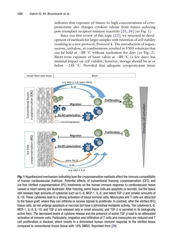
Fig. 1 Hypothesized mechanism indicating how the cryopreservation methods affect the immune compatibility
of human cardiovascular matrices. Potential effects of conventional freezing cryopreservation (CFC) and
ice-free vitrified cryopreservation (IFC) treatments on the human immune response to cardiovascular tissue
(vessel or heart valves) are illustrated. After freezing, some tissue cells are apoptotic or necrotic, but the tissue
still releases high amounts of cytokines such as IL-6, MCP-1, IL-8, and latent TGF-β and smaller amounts of
IL-10. These cytokines lead to a strong activation of blood immune cells. Monocytes and T cells are attracted
to the tissue graft, where they can infiltrate or receive signals to proliferate. In contrast, after the vitrified (IFC)
tissue cells, do not undergo apoptosis or necrosis but have a diminished metabolic activity. The cytokines IL-6,
MCP-1, IL-8, IL-10, and TGF-β are released only in small amounts, and TGF-β is secreted in its biologically
active form. The decreased levels of cytokine release and the presence of active TGF-β lead to an attenuated
activation of immune cells. Particularly, migration and infiltration of T cells and monocytes are reduced and T
cell proliferation is blocked, which results in a diminished human immune response to the vitrified tissue
compared to conventional frozen tissue with 10% DMSO. Reprinted from [24]
596
Kelvin G. M. Brockbank et al.
post-transplant recipient immune reactivity [23, 24] (see Fig. 1).
Since our first review of this topic [12], we returned to development of methods for larger samples with retention of cell viability
resulting in a new protocol, Protocol 4. The introduction of sugars,
sucrose, trehalose, or combinations, resulted in VS55 solutions that
can be held at À80
C without nucleation for days (see Fig. 2).
Short-term exposure of heart valves at À80
C (a few days) has
minimal impact on cell viability; however, storage should be at or
below À135
C. Provided that adequate cryoprotectant tissue
Fig. 1 Hypothesized mechanism indicating how the cryopreservation methods affect the immune compatibility
of human cardiovascular matrices. Potential effects of conventional freezing cryopreservation (CFC) and
ice-free vitrified cryopreservation (IFC) treatments on the human immune response to cardiovascular tissue
(vessel or heart valves) are illustrated. After freezing, some tissue cells are apoptotic or necrotic, but the tissue
still releases high amounts of cytokines such as IL-6, MCP-1, IL-8, and latent TGF-β and smaller amounts of
IL-10. These cytokines lead to a strong activation of blood immune cells. Monocytes and T cells are attracted
to the tissue graft, where they can infiltrate or receive signals to proliferate. In contrast, after the vitrified (IFC)
tissue cells, do not undergo apoptosis or necrosis but have a diminished metabolic activity. The cytokines IL-6,
MCP-1, IL-8, IL-10, and TGF-β are released only in small amounts, and TGF-β is secreted in its biologically
active form. The decreased levels of cytokine release and the presence of active TGF-β lead to an attenuated
activation of immune cells. Particularly, migration and infiltration of T cells and monocytes are reduced and T
cell proliferation is blocked, which results in a diminished human immune response to the vitrified tissue
compared to conventional frozen tissue with 10% DMSO. Reprinted from [24]
596
Kelvin G. M. Brockbank et al.