

and cell structures after cryopreservation and thawing [1, 3, 23,
26]. Cryopreservation of ovarian tissue is performed by a slowfreezing protocol modified from the procedure initially described
by Gosden [27]. In short, ovarian tissue strips (approximately ten
pieces per postpubertal patient with a size of approximately
4 Â 8 Â 1 mm
3 , if a half ovary is laparoscopically removed) were
preincubated for 30 min in 2–4
C pre-cooled sterile-filtered cryopreservation medium containing 10% DMSO. Tissue pieces are
transferred each into one 1.8 mL cryo vial, and the slow-freezing
process is performed in a controlled freezer (e.g., IceCube 14S-A,
SY-LAB, Neupurkersdorf, Austria) which supports automatic
seeding:
1. First remove the stromal tissue/medulla from the cortex with
the aid of Feather precision scalpels no. 22 and Clinastar anatomical forceps. The thickness of the prepared tissue slices
should be approximately 1 mm.
2. After preparation dissect the cortex in pieces of
~8 Â 4 Â 1 mm
3 (according to the age of the patients:
<25 years, smaller pieces with a size of ~5–6 Â 4 Â 1 mm
3 ;
>25 years, bigger pieces of ~8 Â 4 Â 1 mm
3 ).
3. Prepare 2 mm biopsies with a biopsy punch (3 Â 2 mm biopsies
for the fresh viability assay before cryopreservation, after surgical removal and transportation) and additional 3 Â 2 mm biopsies as thawing samples (therefore in total ¼ 6 Â 2 mm
biopsies). The viability assay is a quality control QC measure
to assess primordial follicle density before cryopreservation/
after transportation and after cryopreservation/before transplantation (Fig. 3).
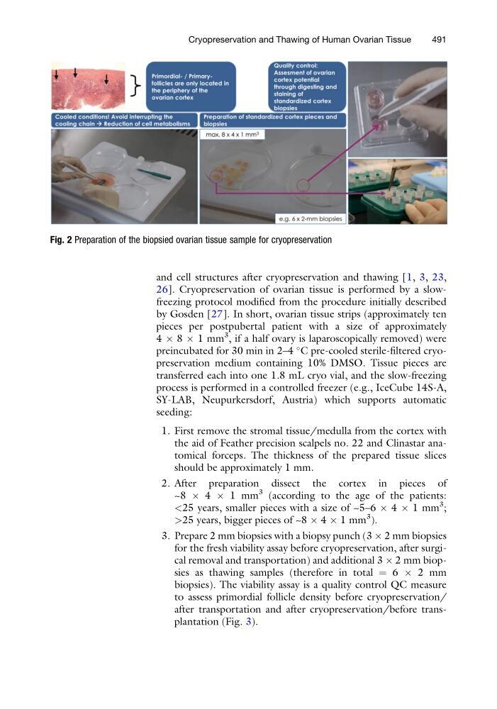
Fig. 2 Preparation of the biopsied ovarian tissue sample for cryopreservation
Cryopreservation and Thawing of Human Ovarian Tissue
491
26]. Cryopreservation of ovarian tissue is performed by a slowfreezing protocol modified from the procedure initially described
by Gosden [27]. In short, ovarian tissue strips (approximately ten
pieces per postpubertal patient with a size of approximately
4 Â 8 Â 1 mm
3 , if a half ovary is laparoscopically removed) were
preincubated for 30 min in 2–4
C pre-cooled sterile-filtered cryopreservation medium containing 10% DMSO. Tissue pieces are
transferred each into one 1.8 mL cryo vial, and the slow-freezing
process is performed in a controlled freezer (e.g., IceCube 14S-A,
SY-LAB, Neupurkersdorf, Austria) which supports automatic
seeding:
1. First remove the stromal tissue/medulla from the cortex with
the aid of Feather precision scalpels no. 22 and Clinastar anatomical forceps. The thickness of the prepared tissue slices
should be approximately 1 mm.
2. After preparation dissect the cortex in pieces of
~8 Â 4 Â 1 mm
3 (according to the age of the patients:
<25 years, smaller pieces with a size of ~5–6 Â 4 Â 1 mm
3 ;
>25 years, bigger pieces of ~8 Â 4 Â 1 mm
3 ).
3. Prepare 2 mm biopsies with a biopsy punch (3 Â 2 mm biopsies
for the fresh viability assay before cryopreservation, after surgical removal and transportation) and additional 3 Â 2 mm biopsies as thawing samples (therefore in total ¼ 6 Â 2 mm
biopsies). The viability assay is a quality control QC measure
to assess primordial follicle density before cryopreservation/
after transportation and after cryopreservation/before transplantation (Fig. 3).
Fig. 2 Preparation of the biopsied ovarian tissue sample for cryopreservation
Cryopreservation and Thawing of Human Ovarian Tissue
491