

4. Prepare aliquots of 32 mL in 50 mL conical centrifuge tubes—
blue caps.
5. Mark the tubes with a label that includes the date of preparation, date of expiration, name of the producer, and composition of the medium with all LOT and REF numbers of
MHM-C and CryoSure-DMSO.
6. Store the aliquots at À20
C. For use on the following day,
thaw one frozen aliquot per patient, by placing overnight at
4
C in a refrigerator.
3.4 Preparation,
Cryopreservation,
and Storage of Ovarian
Tissues
A separate laboratory with a contamination-free environment and a
sterile class II lamina airflow should be available for the preparation
of the ovarian tissue. The laboratory should be accessible via a
separate entrance like an operating theater airlock, and the ovarian
tissue should be prepared in a sterile environment inside a class II
lamina airflow with an integrated cooling plate (e.g., provided by
FRYKA-K€ altetechnik GmbH, Esslingen, Germany). Primordial follicles are only located in the ovarian cortex. Therefore, the tissue is
prepared in such a way that the medulla ovarii is removed from the
cortex ovarii and only a small portion of the residual stroma is left
behind. This makes sure that an optimal starting point for the
neovascularization of the grafts is provided later [25]. Depending
on size and quality, rectangular cortex pieces are cut from the
prepared cortex, equilibrated in a suitable medium for cryopreservation, and then cryopreserved in individual portions for later
therapeutic re-transplantation (see Fig. 2) [1, 3, 23].
The correct selection of the cryoprotectant used is an important factor that is decisive for whether a cryopreservation technique
is successful or not. DMSO, in combination with an appropriate
carrier medium and a protein additive, gives the best survival rates
of the embedded germ cells as well as intact tissue morphologies
Centralized
Cryobank
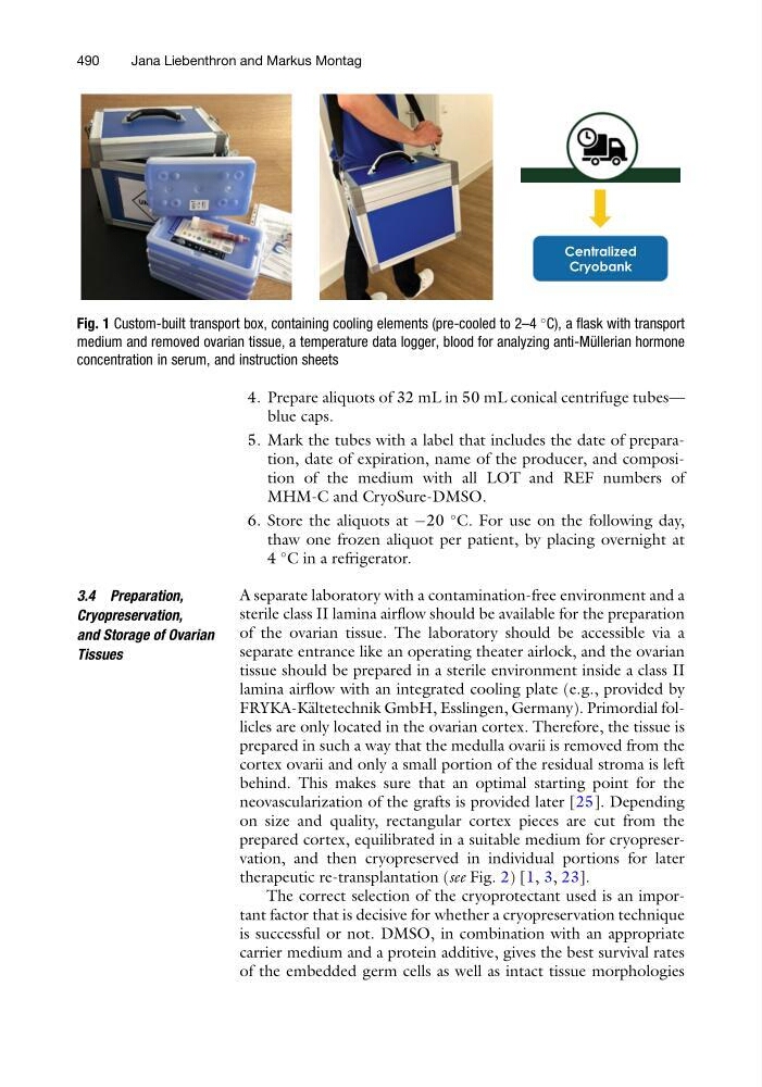
Fig. 1 Custom-built transport box, containing cooling elements (pre-cooled to 2–4
C), a flask with transport
medium and removed ovarian tissue, a temperature data logger, blood for analyzing anti-Mu ¨ llerian hormone
concentration in serum, and instruction sheets
490
Jana Liebenthron and Markus Montag
blue caps.
5. Mark the tubes with a label that includes the date of preparation, date of expiration, name of the producer, and composition of the medium with all LOT and REF numbers of
MHM-C and CryoSure-DMSO.
6. Store the aliquots at À20
C. For use on the following day,
thaw one frozen aliquot per patient, by placing overnight at
4
C in a refrigerator.
3.4 Preparation,
Cryopreservation,
and Storage of Ovarian
Tissues
A separate laboratory with a contamination-free environment and a
sterile class II lamina airflow should be available for the preparation
of the ovarian tissue. The laboratory should be accessible via a
separate entrance like an operating theater airlock, and the ovarian
tissue should be prepared in a sterile environment inside a class II
lamina airflow with an integrated cooling plate (e.g., provided by
FRYKA-K€ altetechnik GmbH, Esslingen, Germany). Primordial follicles are only located in the ovarian cortex. Therefore, the tissue is
prepared in such a way that the medulla ovarii is removed from the
cortex ovarii and only a small portion of the residual stroma is left
behind. This makes sure that an optimal starting point for the
neovascularization of the grafts is provided later [25]. Depending
on size and quality, rectangular cortex pieces are cut from the
prepared cortex, equilibrated in a suitable medium for cryopreservation, and then cryopreserved in individual portions for later
therapeutic re-transplantation (see Fig. 2) [1, 3, 23].
The correct selection of the cryoprotectant used is an important factor that is decisive for whether a cryopreservation technique
is successful or not. DMSO, in combination with an appropriate
carrier medium and a protein additive, gives the best survival rates
of the embedded germ cells as well as intact tissue morphologies
Centralized
Cryobank
Fig. 1 Custom-built transport box, containing cooling elements (pre-cooled to 2–4
C), a flask with transport
medium and removed ovarian tissue, a temperature data logger, blood for analyzing anti-Mu ¨ llerian hormone
concentration in serum, and instruction sheets
490
Jana Liebenthron and Markus Montag