

4. After preparation fill cryopreservation medium (5–7 mL) in
one Oosafe sample container. Transfer the prepared ovarian
tissue pieces and thawing samples (3 Â 2 mm biopsies) for
quality control (QC) into the 5–7 mL cryopreservation
medium.
5. Put the Oosafe sample container with ovarian cortex pieces on
ice/a cooling plate on a shaker inside the class II lamina airflow
for 25 min at 200 cycles/min.
6. Transfer the three remaining 3 Â 2 mm biopsies from different
cortex areas for the fresh viability assay to the four-well Nunc
plate, and start the incubation of the biopsies with 6% CO 2 and
37
C in an incubator (for maximum 2 days/48 h).
7. Pre-cool the Nunc cryo vials and fill afterward cryopreservation
medium (1.7 mL per tube) in each cryo vial inside the class II
lamina airflow. Transfer the ovarian cortex pieces and the samples for QC (thawing samples) into the pre-cooled Nunc cryo
vials (¼ 5 min).
8. Equilibrate the ovarian cortex pieces further on ice, and transfer the tissue in cryo vials to a programmable freezer, such that
they are kept for a total of 10 min at 4
C. The programmed
time in the freezing device is dependent of the distance
between the preparation lab and the freezing device (see
Note 1).
9. Start the freezing program after 40 min (that means: preincubation time for 25 min in Oosafe sample container, 5–10 min
further after transferring into the Nunc cryo vials and 5–10 min
in Nunc cryo vials on ice/or cryo vial cooling container to the
way to the freezing machine—the times in the second to last
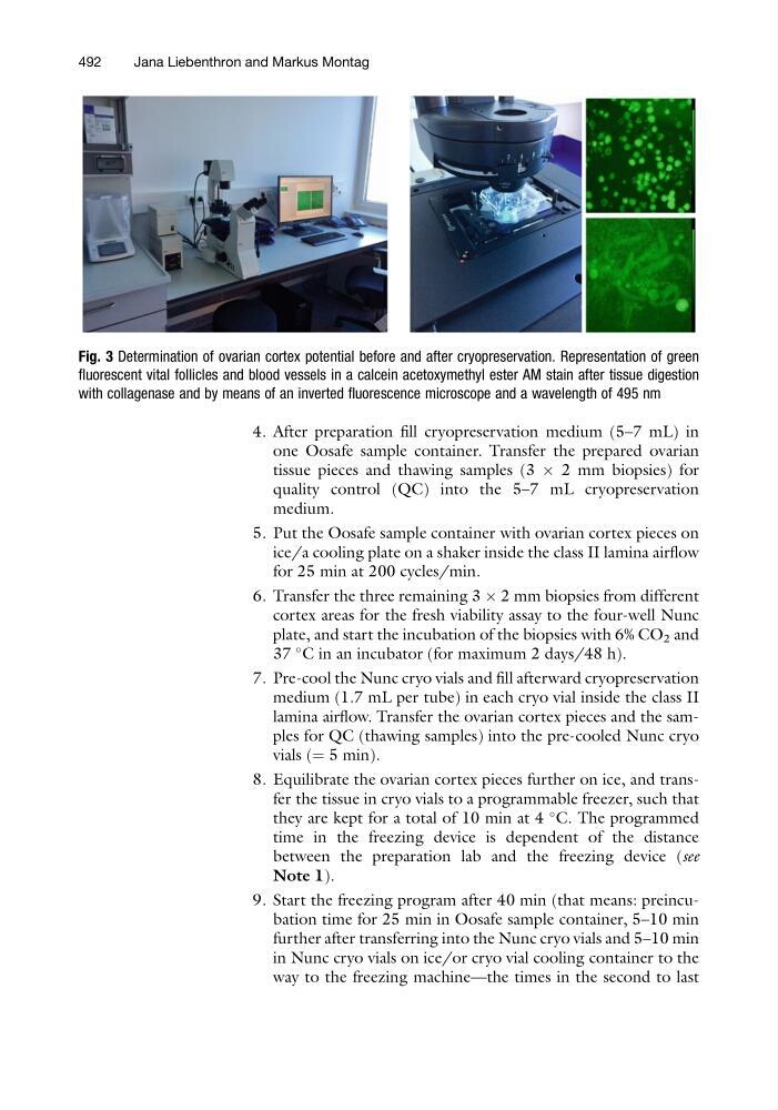
Fig. 3 Determination of ovarian cortex potential before and after cryopreservation. Representation of green
fluorescent vital follicles and blood vessels in a calcein acetoxymethyl ester AM stain after tissue digestion
with collagenase and by means of an inverted fluorescence microscope and a wavelength of 495 nm
492
Jana Liebenthron and Markus Montag
one Oosafe sample container. Transfer the prepared ovarian
tissue pieces and thawing samples (3 Â 2 mm biopsies) for
quality control (QC) into the 5–7 mL cryopreservation
medium.
5. Put the Oosafe sample container with ovarian cortex pieces on
ice/a cooling plate on a shaker inside the class II lamina airflow
for 25 min at 200 cycles/min.
6. Transfer the three remaining 3 Â 2 mm biopsies from different
cortex areas for the fresh viability assay to the four-well Nunc
plate, and start the incubation of the biopsies with 6% CO 2 and
37
C in an incubator (for maximum 2 days/48 h).
7. Pre-cool the Nunc cryo vials and fill afterward cryopreservation
medium (1.7 mL per tube) in each cryo vial inside the class II
lamina airflow. Transfer the ovarian cortex pieces and the samples for QC (thawing samples) into the pre-cooled Nunc cryo
vials (¼ 5 min).
8. Equilibrate the ovarian cortex pieces further on ice, and transfer the tissue in cryo vials to a programmable freezer, such that
they are kept for a total of 10 min at 4
C. The programmed
time in the freezing device is dependent of the distance
between the preparation lab and the freezing device (see
Note 1).
9. Start the freezing program after 40 min (that means: preincubation time for 25 min in Oosafe sample container, 5–10 min
further after transferring into the Nunc cryo vials and 5–10 min
in Nunc cryo vials on ice/or cryo vial cooling container to the
way to the freezing machine—the times in the second to last
Fig. 3 Determination of ovarian cortex potential before and after cryopreservation. Representation of green
fluorescent vital follicles and blood vessels in a calcein acetoxymethyl ester AM stain after tissue digestion
with collagenase and by means of an inverted fluorescence microscope and a wavelength of 495 nm
492
Jana Liebenthron and Markus Montag