

3.90 K 3.0.1.8.
368
15.5 K I.9.7.3.
Biomedical Signal and Image Processing
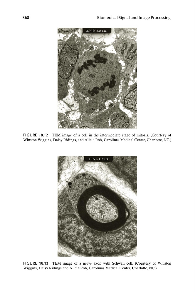
FIGURE 18.12 TEM image of a cell in the intermediate stage of mitosis. (Courtesy of
Winston Wiggins, Daisy Ridings, and Alicia Roh, Carolinas Medical Center, Charlotte, NC.)
FIGURE 18.13 TEM image of a nerve axon with Schwan cell. (Courtesy of Winston
Wiggins, Daisy Ridings and Alicia Roh, Carolinas Medical Center, Charlotte, NC.)
368
15.5 K I.9.7.3.
Biomedical Signal and Image Processing
FIGURE 18.12 TEM image of a cell in the intermediate stage of mitosis. (Courtesy of
Winston Wiggins, Daisy Ridings, and Alicia Roh, Carolinas Medical Center, Charlotte, NC.)
FIGURE 18.13 TEM image of a nerve axon with Schwan cell. (Courtesy of Winston
Wiggins, Daisy Ridings and Alicia Roh, Carolinas Medical Center, Charlotte, NC.)