

367
6.6 1 K H. 5.0.3.
Other Biomedical Imaging Techniques
Two types of electron microscopes can be distinguished: the transmission electron
microscope and the scanning electron microscope. Both types of electron microscopes require a vacuum to minimize the ionization effects of electrons interacting
with air before probing the sample. In addition, the sample preparation will most
certainly result in complete cell death. These requirements rule out in vivo imaging.
18.7.1 TRANSMISSION ELECTRON MICROSCOPY
The transmission electron microscopy (TEM) produces a 2-D attenuation image.
The attenuation images from TEM provide data on the structure of the internal components of the specimen. With special specimen preparation procedures, the TEM
can also be used for the localization of elements, enzymes, and proteins.
The transmission electron microscope operates in a magnification range from
10,000 to 100,000 times with a resolution of approximately 2.5 nm.
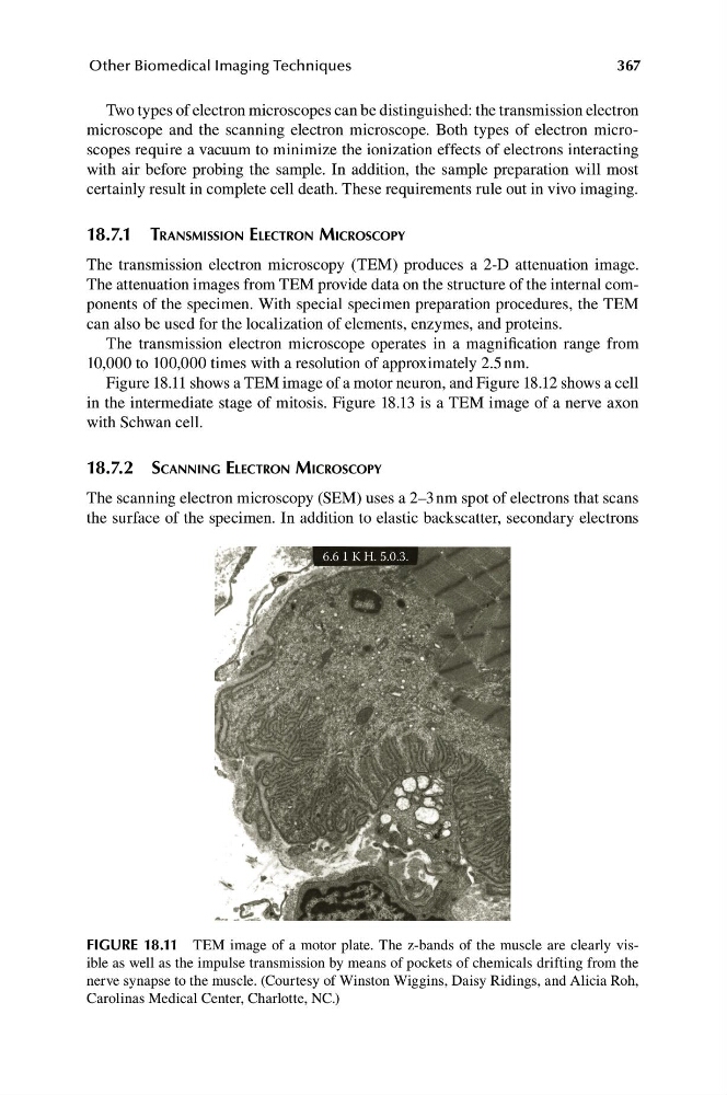
Figure 18.11 shows a TEM image of a motor neuron, and Figure 18.12 shows a cell
in the intermediate stage of mitosis. Figure 18.13 is a TEM image of a nerve axon
with Schwan cell.
18.7.2 SCANNING ELECTRON MICROSCOPY
The scanning electron microscopy (SEM) uses a 2–3 nm spot of electrons that scans
the surface of the specimen. In addition to elastic backscatter, secondary electrons
FIGURE 18.11 TEM image of a motor plate. The z-bands of the muscle are clearly visible as well as the impulse transmission by means of pockets of chemicals drifting from the
nerve synapse to the muscle. (Courtesy of Winston Wiggins, Daisy Ridings, and Alicia Roh,
Carolinas Medical Center, Charlotte, NC.)
6.6 1 K H. 5.0.3.
Other Biomedical Imaging Techniques
Two types of electron microscopes can be distinguished: the transmission electron
microscope and the scanning electron microscope. Both types of electron microscopes require a vacuum to minimize the ionization effects of electrons interacting
with air before probing the sample. In addition, the sample preparation will most
certainly result in complete cell death. These requirements rule out in vivo imaging.
18.7.1 TRANSMISSION ELECTRON MICROSCOPY
The transmission electron microscopy (TEM) produces a 2-D attenuation image.
The attenuation images from TEM provide data on the structure of the internal components of the specimen. With special specimen preparation procedures, the TEM
can also be used for the localization of elements, enzymes, and proteins.
The transmission electron microscope operates in a magnification range from
10,000 to 100,000 times with a resolution of approximately 2.5 nm.
Figure 18.11 shows a TEM image of a motor neuron, and Figure 18.12 shows a cell
in the intermediate stage of mitosis. Figure 18.13 is a TEM image of a nerve axon
with Schwan cell.
18.7.2 SCANNING ELECTRON MICROSCOPY
The scanning electron microscopy (SEM) uses a 2–3 nm spot of electrons that scans
the surface of the specimen. In addition to elastic backscatter, secondary electrons
FIGURE 18.11 TEM image of a motor plate. The z-bands of the muscle are clearly visible as well as the impulse transmission by means of pockets of chemicals drifting from the
nerve synapse to the muscle. (Courtesy of Winston Wiggins, Daisy Ridings, and Alicia Roh,
Carolinas Medical Center, Charlotte, NC.)