

366
Biomedical Signal and Image Processing
1
2
4
3
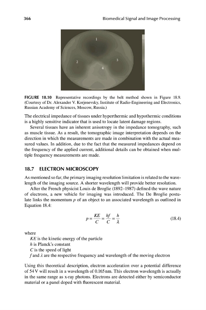
FIGURE 18.10 Representative recordings by the belt method shown in Figure 18.9.
(Courtesy of Dr. Alexander V. Korjenevsky, Institute of Radio-Engineering and Electronics,
Russian Academy of Sciences, Moscow, Russia.)
The electrical impedance of tissues under hyperthermic and hypothermic conditions
is a highly sensitive indicator that is used to locate latent damage regions.
Several tissues have an inherent anisotropy in the impedance tomography, such
as muscle tissue. As a result, the tomographic image interpretation depends on the
direction in which the measurements are made in combination with the actual measured values. In addition, due to the fact that the measured impedances depend on
the frequency of the applied current, additional details can be obtained when multiple frequency measurements are made.
18.7 ELECTRON MICROSCOPY
As mentioned so far, the primary imaging resolution limitation is related to the wavelength of the imaging source. A shorter wavelength will provide better resolution.
After the French physicist Louis de Broglie (1892–1987) defined the wave nature
of electrons, a new vehicle for imaging was introduced. The De Broglie postulate links the momentum p of an object to an associated wavelength as outlined in
Equation 18.4:
KE hf
h
p =
=
=
(18.4)
C
C l
where
KE is the kinetic energy of the particle
h is Planck’s constant
C is the speed of light
f and λ are the respective frequency and wavelength of the moving electron
Using this theoretical description, electron acceleration over a potential difference
of 54 V will result in a wavelength of 0.165 nm. This electron wavelength is actually
in the same range as x-ray photons. Electrons are detected either by semiconductor
material or a panel doped with fluorescent material.
Biomedical Signal and Image Processing
1
2
4
3
FIGURE 18.10 Representative recordings by the belt method shown in Figure 18.9.
(Courtesy of Dr. Alexander V. Korjenevsky, Institute of Radio-Engineering and Electronics,
Russian Academy of Sciences, Moscow, Russia.)
The electrical impedance of tissues under hyperthermic and hypothermic conditions
is a highly sensitive indicator that is used to locate latent damage regions.
Several tissues have an inherent anisotropy in the impedance tomography, such
as muscle tissue. As a result, the tomographic image interpretation depends on the
direction in which the measurements are made in combination with the actual measured values. In addition, due to the fact that the measured impedances depend on
the frequency of the applied current, additional details can be obtained when multiple frequency measurements are made.
18.7 ELECTRON MICROSCOPY
As mentioned so far, the primary imaging resolution limitation is related to the wavelength of the imaging source. A shorter wavelength will provide better resolution.
After the French physicist Louis de Broglie (1892–1987) defined the wave nature
of electrons, a new vehicle for imaging was introduced. The De Broglie postulate links the momentum p of an object to an associated wavelength as outlined in
Equation 18.4:
KE hf
h
p =
=
=
(18.4)
C
C l
where
KE is the kinetic energy of the particle
h is Planck’s constant
C is the speed of light
f and λ are the respective frequency and wavelength of the moving electron
Using this theoretical description, electron acceleration over a potential difference
of 54 V will result in a wavelength of 0.165 nm. This electron wavelength is actually
in the same range as x-ray photons. Electrons are detected either by semiconductor
material or a panel doped with fluorescent material.