

365
Other Biomedical Imaging Techniques
TABLE 18.1
Dielectric Properties of Some
Biological Tissues
Tissue
Resistance (Ω)
Speed of Light
(m/s) × 10 8
Air
High
2.998
Lung
53
0.4206
Fat
113
0.8958
Muscle
50
0.3978
Heart
49.2
0.3912
Cartilage
58
0.4628
Some electrical impedance imaging systems apply tomographic methods to
retrieve depth information from the combined data input from various locations. In
certain cases, the electrodes can be placed in hemispherical or cylindrical symmetric configuration to derive the cross-sectional impedance of an organ or body part;
however, in nonresearch setups, only 2-D surface imaging is performed. Electrical
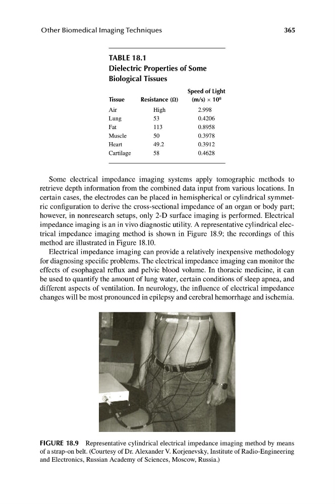
impedance imaging is an in vivo diagnostic utility. A representative cylindrical electrical impedance imaging method is shown in Figure 18.9; the recordings of this
method are illustrated in Figure 18.10.
Electrical impedance imaging can provide a relatively inexpensive methodology
for diagnosing specific problems. The electrical impedance imaging can monitor the
effects of esophageal reflux and pelvic blood volume. In thoracic medicine, it can
be used to quantify the amount of lung water, certain conditions of sleep apnea, and
different aspects of ventilation. In neurology, the influence of electrical impedance
changes will be most pronounced in epilepsy and cerebral hemorrhage and ischemia.
FIGURE 18.9 Representative cylindrical electrical impedance imaging method by means
of a strap-on belt. (Courtesy of Dr. Alexander V. Korjenevsky, Institute of Radio-Engineering
and Electronics, Russian Academy of Sciences, Moscow, Russia.)
Other Biomedical Imaging Techniques
TABLE 18.1
Dielectric Properties of Some
Biological Tissues
Tissue
Resistance (Ω)
Speed of Light
(m/s) × 10 8
Air
High
2.998
Lung
53
0.4206
Fat
113
0.8958
Muscle
50
0.3978
Heart
49.2
0.3912
Cartilage
58
0.4628
Some electrical impedance imaging systems apply tomographic methods to
retrieve depth information from the combined data input from various locations. In
certain cases, the electrodes can be placed in hemispherical or cylindrical symmetric configuration to derive the cross-sectional impedance of an organ or body part;
however, in nonresearch setups, only 2-D surface imaging is performed. Electrical
impedance imaging is an in vivo diagnostic utility. A representative cylindrical electrical impedance imaging method is shown in Figure 18.9; the recordings of this
method are illustrated in Figure 18.10.
Electrical impedance imaging can provide a relatively inexpensive methodology
for diagnosing specific problems. The electrical impedance imaging can monitor the
effects of esophageal reflux and pelvic blood volume. In thoracic medicine, it can
be used to quantify the amount of lung water, certain conditions of sleep apnea, and
different aspects of ventilation. In neurology, the influence of electrical impedance
changes will be most pronounced in epilepsy and cerebral hemorrhage and ischemia.
FIGURE 18.9 Representative cylindrical electrical impedance imaging method by means
of a strap-on belt. (Courtesy of Dr. Alexander V. Korjenevsky, Institute of Radio-Engineering
and Electronics, Russian Academy of Sciences, Moscow, Russia.)