

364
Biomedical Signal and Image Processing
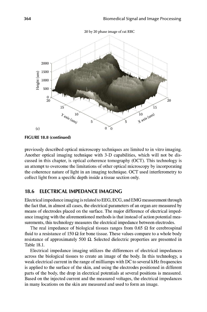
20 by 20 phase image of rat RBC
2000
15
10
5
5
10
15
Y a
X a x is ( u m )
x i s ( u m )
Height (um)
1500
1000
500
0
20
20
(c)
0 0
FIGURE 18.8 (continued)
previously described optical microscopy techniques are limited to in vitro imaging.
Another optical imaging technique with 3-D capabilities, which will not be discussed in this chapter, is optical coherence tomography (OCT). This technology is
an attempt to overcome the limitations of other optical microscopy by incorporating
the coherence nature of light in an imaging technique. OCT used interferometry to
collect light from a specific depth inside a tissue section only.
18.6 ELECTRICAL IMPEDANCE IMAGING
Electrical impedance imaging is related to EEG, ECG, and EMG measurement through
the fact that, in almost all cases, the electrical parameters of an organ are measured by
means of electrodes placed on the surface. The major difference of electrical impedance imaging with the aforementioned methods is that instead of action potential measurements, this technology measures the electrical impedance between electrodes.
The real impedance of biological tissues ranges from 0.65 Ω for cerebrospinal
fluid to a resistance of 150 Ω for bone tissue. These values compare to a whole body
resistance of approximately 500 Ω. Selected dielectric properties are presented in
Table 18.1.
Electrical impedance imaging utilizes the differences of electrical impedances
across the biological tissues to create an image of the body. In this technology, a
weak electrical current in the range of milliamps with DC to several kHz frequencies
is applied to the surface of the skin, and using the electrodes positioned in different
parts of the body, the drop in electrical potentials at several positions is measured.
Based on the injected current and the measured voltages, the electrical impedances
in many locations on the skin are measured and used to form an image.
Biomedical Signal and Image Processing
20 by 20 phase image of rat RBC
2000
15
10
5
5
10
15
Y a
X a x is ( u m )
x i s ( u m )
Height (um)
1500
1000
500
0
20
20
(c)
0 0
FIGURE 18.8 (continued)
previously described optical microscopy techniques are limited to in vitro imaging.
Another optical imaging technique with 3-D capabilities, which will not be discussed in this chapter, is optical coherence tomography (OCT). This technology is
an attempt to overcome the limitations of other optical microscopy by incorporating
the coherence nature of light in an imaging technique. OCT used interferometry to
collect light from a specific depth inside a tissue section only.
18.6 ELECTRICAL IMPEDANCE IMAGING
Electrical impedance imaging is related to EEG, ECG, and EMG measurement through
the fact that, in almost all cases, the electrical parameters of an organ are measured by
means of electrodes placed on the surface. The major difference of electrical impedance imaging with the aforementioned methods is that instead of action potential measurements, this technology measures the electrical impedance between electrodes.
The real impedance of biological tissues ranges from 0.65 Ω for cerebrospinal
fluid to a resistance of 150 Ω for bone tissue. These values compare to a whole body
resistance of approximately 500 Ω. Selected dielectric properties are presented in
Table 18.1.
Electrical impedance imaging utilizes the differences of electrical impedances
across the biological tissues to create an image of the body. In this technology, a
weak electrical current in the range of milliamps with DC to several kHz frequencies
is applied to the surface of the skin, and using the electrodes positioned in different
parts of the body, the drop in electrical potentials at several positions is measured.
Based on the injected current and the measured voltages, the electrical impedances
in many locations on the skin are measured and used to form an image.