

363
5
X a x is ( u m )
x i s ( u m )
Intensity
4.5
15
10
5
5
10
15
X a x i s ( u m )
Y a x is (u m )
Height (au)
4
3.5
3
2.5
2
20
20
(a)
0 0
Topography of rat RBC
800
8
6
4
2
2
4
6
8
Y a
Height (nm)
600
400
200
0
10
10
(b)
0 0
Other Biomedical Imaging Techniques
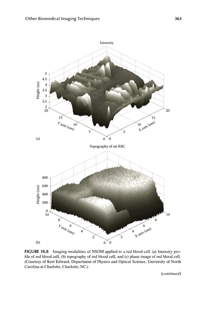
FIGURE 18.8 Imaging modalities of NSOM applied to a red blood cell. (a) Intensity profile of red blood cell, (b) topography of red blood cell, and (c) phase image of red blood cell.
(Courtesy of Kert Edward, Department of Physics and Optical Science, University of North
Carolina at Charlotte, Charlotte, NC.)
(continued)
5
X a x is ( u m )
x i s ( u m )
Intensity
4.5
15
10
5
5
10
15
X a x i s ( u m )
Y a x is (u m )
Height (au)
4
3.5
3
2.5
2
20
20
(a)
0 0
Topography of rat RBC
800
8
6
4
2
2
4
6
8
Y a
Height (nm)
600
400
200
0
10
10
(b)
0 0
Other Biomedical Imaging Techniques
FIGURE 18.8 Imaging modalities of NSOM applied to a red blood cell. (a) Intensity profile of red blood cell, (b) topography of red blood cell, and (c) phase image of red blood cell.
(Courtesy of Kert Edward, Department of Physics and Optical Science, University of North
Carolina at Charlotte, Charlotte, NC.)
(continued)