

360
Biomedical Signal and Image Processing
Indirect functional imaging of the cell is possible using fluorescent microscopy.
This is done by monitoring the oxygen level in different parts of a sample. In order
to do so, an oxygen-quenching fluorescent dye, tris (1,10-phenanthroline) ruthenium
(II) chloral hydrate, incorporated in a silicone rubber membrane is attached to an
excised portion of the circulation system. The contact between the membrane and
the vasculature then indicates the oxygen concentration and therefore the distribution of the blood flowing through the respective blood vessels. The membrane
fluoresces proportional to the oxygen concentration of the blood in contact with the
membrane.
The image acquisition for the fluorescent microscopy will be identical to that for
the standard optical microscope. The only difference will be the lack of specific
anatomical details due to the optical filtering. It is therefore customary to combine
standard optical microscopy with fluorescent microscopy.
Images created by regular optical as well as fluorescent microscopy are twodimensional (2-D) and therefore miss the details on the third dimension. The need
for examining the third dimension of the samples led to the development of yet
another imaging technique based on the principles of the fluorescent microscope.
The next section describes this technology that is called confocal microscopy.
18.4 CONFOCAL MICROSCOPY
Confocal microscopy is a “stereo” version of fluoroscopic microscopy with the
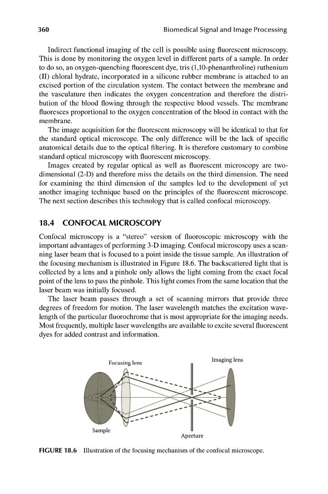
important advantages of performing 3-D imaging. Confocal microscopy uses a scanning laser beam that is focused to a point inside the tissue sample. An illustration of
the focusing mechanism is illustrated in Figure 18.6. The backscattered light that is
collected by a lens and a pinhole only allows the light coming from the exact focal
point of the lens to pass the pinhole. This light comes from the same location that the
laser beam was initially focused.
The laser beam passes through a set of scanning mirrors that provide three
degrees of freedom for motion. The laser wavelength matches the excitation wavelength of the particular fluorochrome that is most appropriate for the imaging needs.
Most frequently, multiple laser wavelengths are available to excite several fluorescent
dyes for added contrast and information.
Imaging lens
Focusing lens
Sample
Aperture
FIGURE 18.6 Illustration of the focusing mechanism of the confocal microscope.
Biomedical Signal and Image Processing
Indirect functional imaging of the cell is possible using fluorescent microscopy.
This is done by monitoring the oxygen level in different parts of a sample. In order
to do so, an oxygen-quenching fluorescent dye, tris (1,10-phenanthroline) ruthenium
(II) chloral hydrate, incorporated in a silicone rubber membrane is attached to an
excised portion of the circulation system. The contact between the membrane and
the vasculature then indicates the oxygen concentration and therefore the distribution of the blood flowing through the respective blood vessels. The membrane
fluoresces proportional to the oxygen concentration of the blood in contact with the
membrane.
The image acquisition for the fluorescent microscopy will be identical to that for
the standard optical microscope. The only difference will be the lack of specific
anatomical details due to the optical filtering. It is therefore customary to combine
standard optical microscopy with fluorescent microscopy.
Images created by regular optical as well as fluorescent microscopy are twodimensional (2-D) and therefore miss the details on the third dimension. The need
for examining the third dimension of the samples led to the development of yet
another imaging technique based on the principles of the fluorescent microscope.
The next section describes this technology that is called confocal microscopy.
18.4 CONFOCAL MICROSCOPY
Confocal microscopy is a “stereo” version of fluoroscopic microscopy with the
important advantages of performing 3-D imaging. Confocal microscopy uses a scanning laser beam that is focused to a point inside the tissue sample. An illustration of
the focusing mechanism is illustrated in Figure 18.6. The backscattered light that is
collected by a lens and a pinhole only allows the light coming from the exact focal
point of the lens to pass the pinhole. This light comes from the same location that the
laser beam was initially focused.
The laser beam passes through a set of scanning mirrors that provide three
degrees of freedom for motion. The laser wavelength matches the excitation wavelength of the particular fluorochrome that is most appropriate for the imaging needs.
Most frequently, multiple laser wavelengths are available to excite several fluorescent
dyes for added contrast and information.
Imaging lens
Focusing lens
Sample
Aperture
FIGURE 18.6 Illustration of the focusing mechanism of the confocal microscope.