

361
Other Biomedical Imaging Techniques
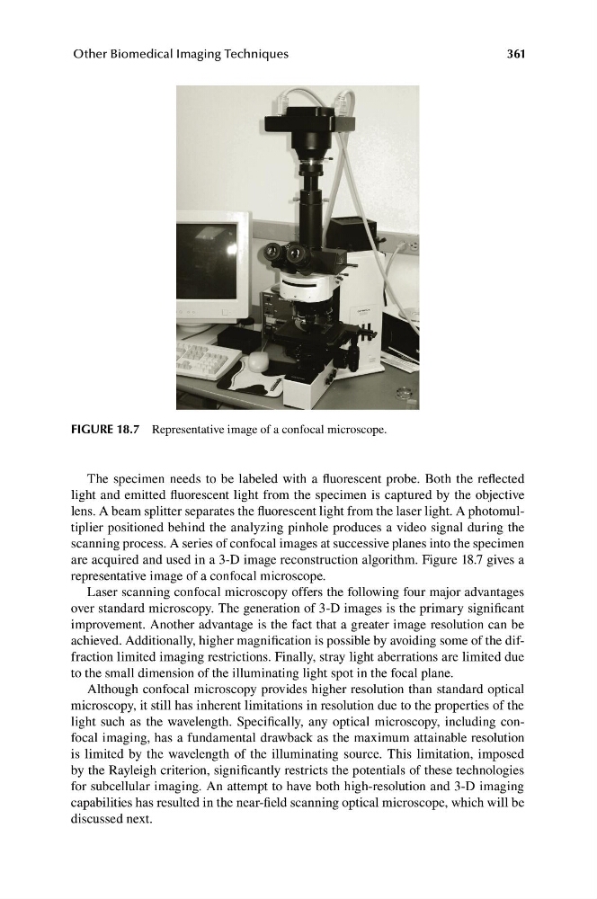
FIGURE 18.7 Representative image of a confocal microscope.
The specimen needs to be labeled with a fluorescent probe. Both the reflected
light and emitted fluorescent light from the specimen is captured by the objective
lens. A beam splitter separates the fluorescent light from the laser light. A photomultiplier positioned behind the analyzing pinhole produces a video signal during the
scanning process. A series of confocal images at successive planes into the specimen
are acquired and used in a 3-D image reconstruction algorithm. Figure 18.7 gives a
representative image of a confocal microscope.
Laser scanning confocal microscopy offers the following four major advantages
over standard microscopy. The generation of 3-D images is the primary significant
improvement. Another advantage is the fact that a greater image resolution can be
achieved. Additionally, higher magnification is possible by avoiding some of the diffraction limited imaging restrictions. Finally, stray light aberrations are limited due
to the small dimension of the illuminating light spot in the focal plane.
Although confocal microscopy provides higher resolution than standard optical
microscopy, it still has inherent limitations in resolution due to the properties of the
light such as the wavelength. Specifically, any optical microscopy, including confocal imaging, has a fundamental drawback as the maximum attainable resolution
is limited by the wavelength of the illuminating source. This limitation, imposed
by the Rayleigh criterion, significantly restricts the potentials of these technologies
for subcellular imaging. An attempt to have both high-resolution and 3-D imaging
capabilities has resulted in the near-field scanning optical microscope, which will be
discussed next.
Other Biomedical Imaging Techniques
FIGURE 18.7 Representative image of a confocal microscope.
The specimen needs to be labeled with a fluorescent probe. Both the reflected
light and emitted fluorescent light from the specimen is captured by the objective
lens. A beam splitter separates the fluorescent light from the laser light. A photomultiplier positioned behind the analyzing pinhole produces a video signal during the
scanning process. A series of confocal images at successive planes into the specimen
are acquired and used in a 3-D image reconstruction algorithm. Figure 18.7 gives a
representative image of a confocal microscope.
Laser scanning confocal microscopy offers the following four major advantages
over standard microscopy. The generation of 3-D images is the primary significant
improvement. Another advantage is the fact that a greater image resolution can be
achieved. Additionally, higher magnification is possible by avoiding some of the diffraction limited imaging restrictions. Finally, stray light aberrations are limited due
to the small dimension of the illuminating light spot in the focal plane.
Although confocal microscopy provides higher resolution than standard optical
microscopy, it still has inherent limitations in resolution due to the properties of the
light such as the wavelength. Specifically, any optical microscopy, including confocal imaging, has a fundamental drawback as the maximum attainable resolution
is limited by the wavelength of the illuminating source. This limitation, imposed
by the Rayleigh criterion, significantly restricts the potentials of these technologies
for subcellular imaging. An attempt to have both high-resolution and 3-D imaging
capabilities has resulted in the near-field scanning optical microscope, which will be
discussed next.