

359
Other Biomedical Imaging Techniques
FIGURE 18.4 Picture of a fluorescent microscope with red, green, and blue lasers attached.
and rhodamine when excited by green–yellow light emits deep red. The distribution
of different molecules within one single cell can be visualized by coupling one tag
molecule to fluorescence and another to rhodamine. The two tags can be identified by switching the optical filters that are transparent at the respective fluorescent
wavelengths. Figure 18.5 shows the histology of a mouse uterus stained by the stain
phycoerythrin (PE) that emits red light under green illumination.
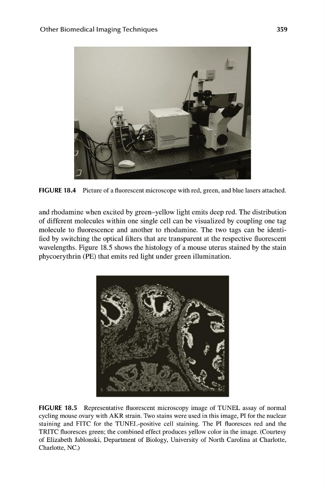
FIGURE 18.5 Representative fluorescent microscopy image of TUNEL assay of normal
cycling mouse ovary with AKR strain. Two stains were used in this image, PI for the nuclear
staining and FITC for the TUNEL-positive cell staining. The PI fluoresces red and the
TRITC fluoresces green; the combined effect produces yellow color in the image. (Courtesy
of Elizabeth Jablonski, Department of Biology, University of North Carolina at Charlotte,
Charlotte, NC.)
Other Biomedical Imaging Techniques
FIGURE 18.4 Picture of a fluorescent microscope with red, green, and blue lasers attached.
and rhodamine when excited by green–yellow light emits deep red. The distribution
of different molecules within one single cell can be visualized by coupling one tag
molecule to fluorescence and another to rhodamine. The two tags can be identified by switching the optical filters that are transparent at the respective fluorescent
wavelengths. Figure 18.5 shows the histology of a mouse uterus stained by the stain
phycoerythrin (PE) that emits red light under green illumination.
FIGURE 18.5 Representative fluorescent microscopy image of TUNEL assay of normal
cycling mouse ovary with AKR strain. Two stains were used in this image, PI for the nuclear
staining and FITC for the TUNEL-positive cell staining. The PI fluoresces red and the
TRITC fluoresces green; the combined effect produces yellow color in the image. (Courtesy
of Elizabeth Jablonski, Department of Biology, University of North Carolina at Charlotte,
Charlotte, NC.)