

358
Biomedical Signal and Image Processing
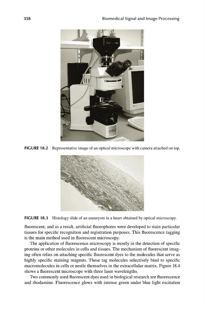
FIGURE 18.2 Representative image of an optical microscope with camera attached on top.
FIGURE 18.3 Histology slide of an aneurysm in a heart obtained by optical microscopy.
fluorescent, and as a result, artificial fluorophores were developed to stain particular
tissues for specific recognition and registration purposes. This fluorescence tagging
is the main method used in fluorescent microscopy.
The application of fluorescence microscopy is mostly in the detection of specific
proteins or other molecules in cells and tissues. The mechanism of fluorescent imaging often relies on attaching specific fluorescent dyes to the molecules that serve as
highly specific staining reagents. These tag molecules selectively bind to specific
macromolecules in cells or nestle themselves in the extracellular matrix. Figure 18.4
shows a fluorescent microscope with three laser wavelengths.
Two commonly used fluorescent dyes used in biological research are fluorescence
and rhodamine. Fluorescence glows with intense green under blue light excitation
Biomedical Signal and Image Processing
FIGURE 18.2 Representative image of an optical microscope with camera attached on top.
FIGURE 18.3 Histology slide of an aneurysm in a heart obtained by optical microscopy.
fluorescent, and as a result, artificial fluorophores were developed to stain particular
tissues for specific recognition and registration purposes. This fluorescence tagging
is the main method used in fluorescent microscopy.
The application of fluorescence microscopy is mostly in the detection of specific
proteins or other molecules in cells and tissues. The mechanism of fluorescent imaging often relies on attaching specific fluorescent dyes to the molecules that serve as
highly specific staining reagents. These tag molecules selectively bind to specific
macromolecules in cells or nestle themselves in the extracellular matrix. Figure 18.4
shows a fluorescent microscope with three laser wavelengths.
Two commonly used fluorescent dyes used in biological research are fluorescence
and rhodamine. Fluorescence glows with intense green under blue light excitation