

349
Positron Emission Tomography
dementia forms the justification for the use of PET imaging. PET imaging shows the
metabolic degeneration of the neurotransmitters in the brain of an Alzheimer patient.
PET can track the different stages of reduced brain function. While in the early
stages of Alzheimer’s disease, only limited areas of the brain will be detected to have
a lower level of function, in the later stages of Alzheimer’s disease, the metabolic
activity of larger areas of the brain will progressively appear affected. Particularly,
in Alzheimer’s disease, the disease follows a certain pattern in affecting the brain
under PET imaging. The disease pattern can often be recognized several years in
advance of the manifestation of episodes of confusion and recognizable category of
dementia or depression.
Other brain disorders that can be located by PET scans are Parkinson’s disease
and schizophrenia. Other neurological diagnoses that can be performed using PET
include the diagnostics and study of the brain activities in epilepsy and stroke. For
instance, PET can be used to determine the location of epileptic seizures prior to
surgery to develop a road map for the procedure.
Considering the imaging of the neural structures, the pivotal advantage of PET
over many of the other available imaging modalities is the ability to reveal activity of
neuroreceptors such as the ones that use the neurotransmitters serotonin, dopamine,
and noradrenaline. Typical MRI imaging systems are unable to identify neurochemical sites due to the low neurotransmitter concentrations involved (in the order of
micromolar concentrations).
17.6.3 FUNCTIONAL HEART IMAGING
For diagnostic applications in cardiology, PET with FDG is used to functionally image
the heart tissue after a heart attack and determine if there is any latent damage in the
heart muscle. In the diagnosis of heart disease, the dead tissue can be separated from
the living tissue in a PET scan based on the oxygen isotope interaction. PET imaging is
also useful in predicting the success of angioplasty or even bypass surgery. In another
cardiovascular application, PET scanning is used to determine blockage of coronary
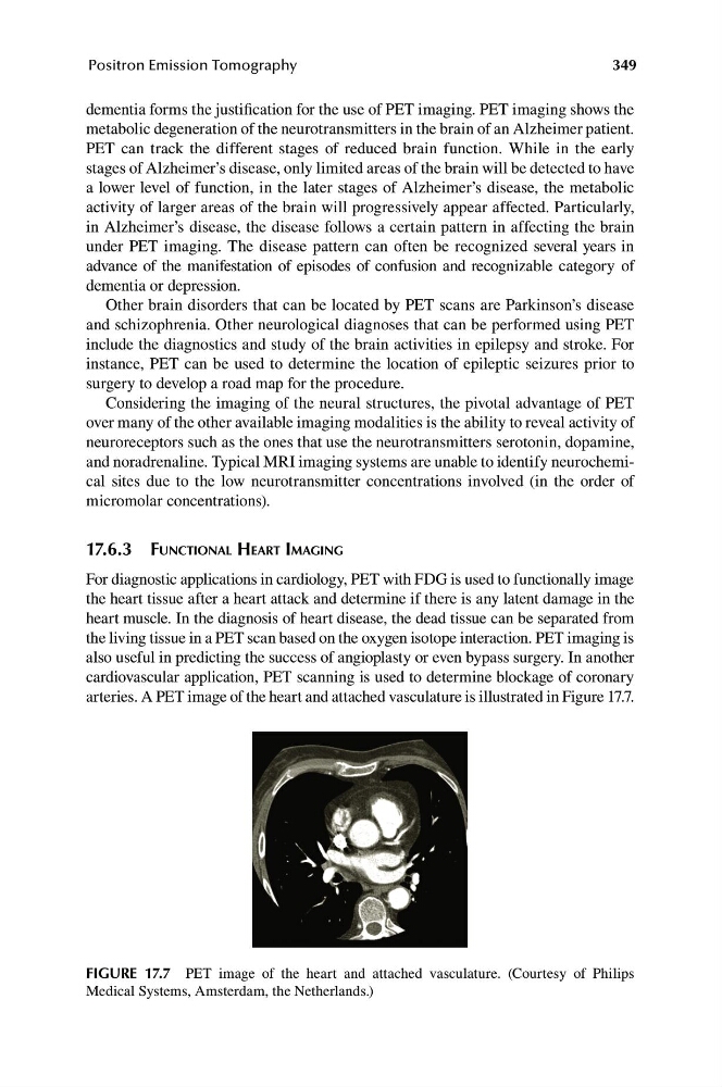
arteries. A PET image of the heart and attached vasculature is illustrated in Figure 17.7.
FIGURE 17.7 PET image of the heart and attached vasculature. (Courtesy of Philips
Medical Systems, Amsterdam, the Netherlands.)
Positron Emission Tomography
dementia forms the justification for the use of PET imaging. PET imaging shows the
metabolic degeneration of the neurotransmitters in the brain of an Alzheimer patient.
PET can track the different stages of reduced brain function. While in the early
stages of Alzheimer’s disease, only limited areas of the brain will be detected to have
a lower level of function, in the later stages of Alzheimer’s disease, the metabolic
activity of larger areas of the brain will progressively appear affected. Particularly,
in Alzheimer’s disease, the disease follows a certain pattern in affecting the brain
under PET imaging. The disease pattern can often be recognized several years in
advance of the manifestation of episodes of confusion and recognizable category of
dementia or depression.
Other brain disorders that can be located by PET scans are Parkinson’s disease
and schizophrenia. Other neurological diagnoses that can be performed using PET
include the diagnostics and study of the brain activities in epilepsy and stroke. For
instance, PET can be used to determine the location of epileptic seizures prior to
surgery to develop a road map for the procedure.
Considering the imaging of the neural structures, the pivotal advantage of PET
over many of the other available imaging modalities is the ability to reveal activity of
neuroreceptors such as the ones that use the neurotransmitters serotonin, dopamine,
and noradrenaline. Typical MRI imaging systems are unable to identify neurochemical sites due to the low neurotransmitter concentrations involved (in the order of
micromolar concentrations).
17.6.3 FUNCTIONAL HEART IMAGING
For diagnostic applications in cardiology, PET with FDG is used to functionally image
the heart tissue after a heart attack and determine if there is any latent damage in the
heart muscle. In the diagnosis of heart disease, the dead tissue can be separated from
the living tissue in a PET scan based on the oxygen isotope interaction. PET imaging is
also useful in predicting the success of angioplasty or even bypass surgery. In another
cardiovascular application, PET scanning is used to determine blockage of coronary
arteries. A PET image of the heart and attached vasculature is illustrated in Figure 17.7.
FIGURE 17.7 PET image of the heart and attached vasculature. (Courtesy of Philips
Medical Systems, Amsterdam, the Netherlands.)