

1
222–234 1
230–242 1
238–250 1
246–258 1
254–266 1
262–274 1
270–282
1
278–290 1
286–298 1
294–306 1
302–314 1
310–322 1
318–330 1
325–338
1
334–346 1
342–354 1
350–362 1
358–370 1
366–378 1
374–386 1
382–394
348
Biomedical Signal and Image Processing
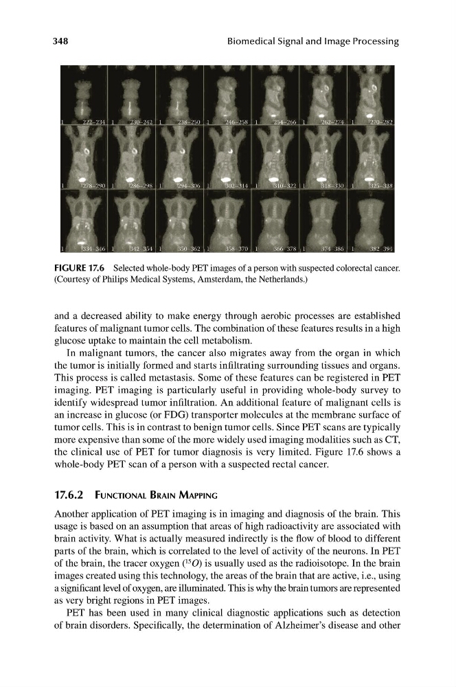
FIGURE 17.6 Selected whole-body PET images of a person with suspected colorectal cancer.
(Courtesy of Philips Medical Systems, Amsterdam, the Netherlands.)
and a decreased ability to make energy through aerobic processes are established
features of malignant tumor cells. The combination of these features results in a high
glucose uptake to maintain the cell metabolism.
In malignant tumors, the cancer also migrates away from the organ in which
the tumor is initially formed and starts infiltrating surrounding tissues and organs.
This process is called metastasis. Some of these features can be registered in PET
imaging. PET imaging is particularly useful in providing whole-body survey to
identify widespread tumor infiltration. An additional feature of malignant cells is
an increase in glucose (or FDG) transporter molecules at the membrane surface of
tumor cells. This is in contrast to benign tumor cells. Since PET scans are typically
more expensive than some of the more widely used imaging modalities such as CT,
the clinical use of PET for tumor diagnosis is very limited. Figure 17.6 shows a
whole-body PET scan of a person with a suspected rectal cancer.
17.6.2 FUNCTIONAL BRAIN MAPPING
Another application of PET imaging is in imaging and diagnosis of the brain. This
usage is based on an assumption that areas of high radioactivity are associated with
brain activity. What is actually measured indirectly is the flow of blood to different
parts of the brain, which is correlated to the level of activity of the neurons. In PET
of the brain, the tracer oxygen ( 15 O) is usually used as the radioisotope. In the brain
images created using this technology, the areas of the brain that are active, i.e., using
a significant level of oxygen, are illuminated. This is why the brain tumors are represented
as very bright regions in PET images.
PET has been used in many clinical diagnostic applications such as detection
of brain disorders. Specifically, the determination of Alzheimer’s disease and other
222–234 1
230–242 1
238–250 1
246–258 1
254–266 1
262–274 1
270–282
1
278–290 1
286–298 1
294–306 1
302–314 1
310–322 1
318–330 1
325–338
1
334–346 1
342–354 1
350–362 1
358–370 1
366–378 1
374–386 1
382–394
348
Biomedical Signal and Image Processing
FIGURE 17.6 Selected whole-body PET images of a person with suspected colorectal cancer.
(Courtesy of Philips Medical Systems, Amsterdam, the Netherlands.)
and a decreased ability to make energy through aerobic processes are established
features of malignant tumor cells. The combination of these features results in a high
glucose uptake to maintain the cell metabolism.
In malignant tumors, the cancer also migrates away from the organ in which
the tumor is initially formed and starts infiltrating surrounding tissues and organs.
This process is called metastasis. Some of these features can be registered in PET
imaging. PET imaging is particularly useful in providing whole-body survey to
identify widespread tumor infiltration. An additional feature of malignant cells is
an increase in glucose (or FDG) transporter molecules at the membrane surface of
tumor cells. This is in contrast to benign tumor cells. Since PET scans are typically
more expensive than some of the more widely used imaging modalities such as CT,
the clinical use of PET for tumor diagnosis is very limited. Figure 17.6 shows a
whole-body PET scan of a person with a suspected rectal cancer.
17.6.2 FUNCTIONAL BRAIN MAPPING
Another application of PET imaging is in imaging and diagnosis of the brain. This
usage is based on an assumption that areas of high radioactivity are associated with
brain activity. What is actually measured indirectly is the flow of blood to different
parts of the brain, which is correlated to the level of activity of the neurons. In PET
of the brain, the tracer oxygen ( 15 O) is usually used as the radioisotope. In the brain
images created using this technology, the areas of the brain that are active, i.e., using
a significant level of oxygen, are illuminated. This is why the brain tumors are represented
as very bright regions in PET images.
PET has been used in many clinical diagnostic applications such as detection
of brain disorders. Specifically, the determination of Alzheimer’s disease and other