

350
Table
Biomedical Signal and Image Processing
Despite the numerous benefits of PET for cardiovascular imaging, often the relatively inexpensive SPECT is used for this purpose. SPECT, like PET, acquires the concentration distribution of radionucleotides in a patient’s body. The major differences
between PET and SPECT are in the choice of radioisotopes and hence the energy of
the emission. In PET, two gamma photons are created from the emitted positron particle, while, in SPECT, only one single photon is emitted without a go-between with considerably less energy, approximately 140 keV compared to 511 keV. Due to the nature
and configuration of the single photon emitted from the radionucleotides in SPECT,
special collimators are required to acquire an image from multiple angles. The use
of collimators dramatically reduces the detection efficiency in comparison with PET.
This is in sharp contrast with PET imaging, which relies on the perpendicular nature
of the emitted gamma photons of annihilation of the positron emitted from the isotope.
Generally, PET has two orders of magnitude greater number of detectors than
SPECT, giving PET imaging a much higher resolution. The advantage of SPECT is
the wide variety of radionucleotides that are available and the resulting bigger range
of detection of diseases. The cost of SPECT imaging is approximately one-third that
of PET, which gives it an advantage over PET in certain cases. However, the lower
resolution remains an obstacle. SPECT will be described in more detail in Chapter 18.
17.6.4 ANATOMICAL IMAGING
As previously discussed, PET provides no significant anatomical information primarily due to the fact that the uptake of radioisotopes in some tissues such as bone
is too slow to be recorded. For instance, due to the half-life of the isotopes and the
assembly time for bone tissue, the radioactivity has dropped below the detection
level at the time of incorporation in the skeletal system.
The anatomical features that can be recognized in PET are mainly due to the a
priori metabolic activities that are well known for certain organs such as in the brain
and the heart. Other anatomical features will need to be resolved by registering
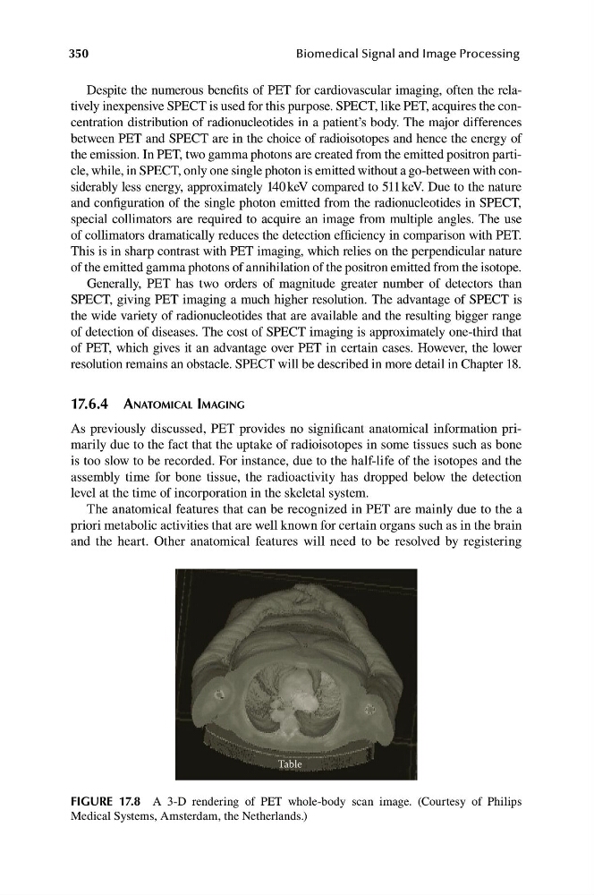
FIGURE 17.8 A 3-D rendering of PET whole-body scan image. (Courtesy of Philips
Medical Systems, Amsterdam, the Netherlands.)
Table
Biomedical Signal and Image Processing
Despite the numerous benefits of PET for cardiovascular imaging, often the relatively inexpensive SPECT is used for this purpose. SPECT, like PET, acquires the concentration distribution of radionucleotides in a patient’s body. The major differences
between PET and SPECT are in the choice of radioisotopes and hence the energy of
the emission. In PET, two gamma photons are created from the emitted positron particle, while, in SPECT, only one single photon is emitted without a go-between with considerably less energy, approximately 140 keV compared to 511 keV. Due to the nature
and configuration of the single photon emitted from the radionucleotides in SPECT,
special collimators are required to acquire an image from multiple angles. The use
of collimators dramatically reduces the detection efficiency in comparison with PET.
This is in sharp contrast with PET imaging, which relies on the perpendicular nature
of the emitted gamma photons of annihilation of the positron emitted from the isotope.
Generally, PET has two orders of magnitude greater number of detectors than
SPECT, giving PET imaging a much higher resolution. The advantage of SPECT is
the wide variety of radionucleotides that are available and the resulting bigger range
of detection of diseases. The cost of SPECT imaging is approximately one-third that
of PET, which gives it an advantage over PET in certain cases. However, the lower
resolution remains an obstacle. SPECT will be described in more detail in Chapter 18.
17.6.4 ANATOMICAL IMAGING
As previously discussed, PET provides no significant anatomical information primarily due to the fact that the uptake of radioisotopes in some tissues such as bone
is too slow to be recorded. For instance, due to the half-life of the isotopes and the
assembly time for bone tissue, the radioactivity has dropped below the detection
level at the time of incorporation in the skeletal system.
The anatomical features that can be recognized in PET are mainly due to the a
priori metabolic activities that are well known for certain organs such as in the brain
and the heart. Other anatomical features will need to be resolved by registering
FIGURE 17.8 A 3-D rendering of PET whole-body scan image. (Courtesy of Philips
Medical Systems, Amsterdam, the Netherlands.)