

343
Positron Emission Tomography
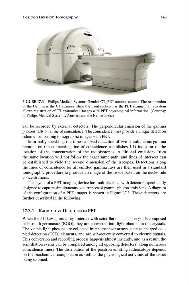
FIGURE 17.3 Philips Medical Systems Gemini CT_PET combo scanner. The rear section
of the Gemini is the CT scanner while the front section has the PET scanner. This system
allows registration of CT anatomical images with PET physiological information. (Courtesy
of Philips Medical Systems, Amsterdam, the Netherlands.)
can be recorded by external detectors. The perpendicular emission of the gamma
photons falls on a line of coincidence. The coincidence lines provide a unique detection
scheme for forming tomographic images with PET.
Informally speaking, the time-resolved detection of two simultaneous gamma
photons on the connecting line of coincidence establishes 1-D indicator of the
location of the concentration of the radioisotopes. Additional emissions from
the same location will not follow the exact same path, and lines of intersect can
be established to yield the second dimension of the isotopes. Detections along
the lines of coincidence for all emitted gamma rays are then used in a standard
tomographic procedure to produce an image of the tissue based on the nucleotide
concentrations.
The layout of a PET imaging device has multiple rings with detectors specifically
designed to capture simultaneous occurrences of gamma photon emissions. A diagram
of the configuration of a PET imager is shown in Figure 17.3. These detectors are
further described in the following.
17.3.1 RADIOACTIVE DETECTION IN PET
When the 511 keV gamma rays interact with scintillation such as crystals composed
of bismuth germanate (BGO), they are converted into light photons in the crystals.
The visible light photons are collected by photosensor arrays, such as charged coupled detection (CCD) elements, and are subsequently converted to electric signals.
This conversion and recording process happens almost instantly, and as a result, the
scintillation events can be compared among all opposing detectors (along numerous
coincidence lines). The distribution of the positron emitting radioisotope depends
on the biochemical composition as well as the physiological activities of the tissue
being scanned.
Positron Emission Tomography
FIGURE 17.3 Philips Medical Systems Gemini CT_PET combo scanner. The rear section
of the Gemini is the CT scanner while the front section has the PET scanner. This system
allows registration of CT anatomical images with PET physiological information. (Courtesy
of Philips Medical Systems, Amsterdam, the Netherlands.)
can be recorded by external detectors. The perpendicular emission of the gamma
photons falls on a line of coincidence. The coincidence lines provide a unique detection
scheme for forming tomographic images with PET.
Informally speaking, the time-resolved detection of two simultaneous gamma
photons on the connecting line of coincidence establishes 1-D indicator of the
location of the concentration of the radioisotopes. Additional emissions from
the same location will not follow the exact same path, and lines of intersect can
be established to yield the second dimension of the isotopes. Detections along
the lines of coincidence for all emitted gamma rays are then used in a standard
tomographic procedure to produce an image of the tissue based on the nucleotide
concentrations.
The layout of a PET imaging device has multiple rings with detectors specifically
designed to capture simultaneous occurrences of gamma photon emissions. A diagram
of the configuration of a PET imager is shown in Figure 17.3. These detectors are
further described in the following.
17.3.1 RADIOACTIVE DETECTION IN PET
When the 511 keV gamma rays interact with scintillation such as crystals composed
of bismuth germanate (BGO), they are converted into light photons in the crystals.
The visible light photons are collected by photosensor arrays, such as charged coupled detection (CCD) elements, and are subsequently converted to electric signals.
This conversion and recording process happens almost instantly, and as a result, the
scintillation events can be compared among all opposing detectors (along numerous
coincidence lines). The distribution of the positron emitting radioisotope depends
on the biochemical composition as well as the physiological activities of the tissue
being scanned.