

342
Biomedical Signal and Image Processing
17.3 PET SIGNAL ACQUISITION
All short-lived radioisotopes used in PET imaging decay by positron emission.
Positrons (β+) are emitted from the nucleus of radioisotopes that are unstable because
they have an excessive number of protons and a positive charge. The positron emission is different from the free proton or odd proton count utilized in NMR imaging
as described in Chapter 15.
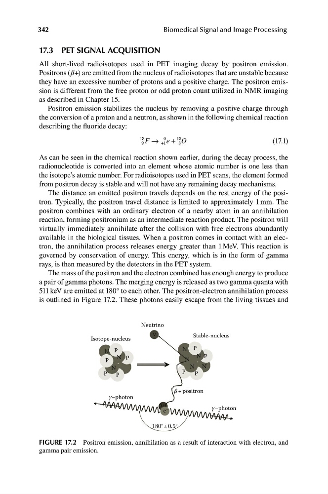
Positron emission stabilizes the nucleus by removing a positive charge through
the conversion of a proton and a neutron, as shown in the following chemical reaction
describing the fluoride decay:
18
0
18
9 F → +1 e + 8 O
(17.1)
As can be seen in the chemical reaction shown earlier, during the decay process, the
radionucleotide is converted into an element whose atomic number is one less than
the isotope’s atomic number. For radioisotopes used in PET scans, the element formed
from positron decay is stable and will not have any remaining decay mechanisms.
The distance an emitted positron travels depends on the rest energy of the positron. Typically, the positron travel distance is limited to approximately 1 mm. The
positron combines with an ordinary electron of a nearby atom in an annihilation
reaction, forming positronium as an intermediate reaction product. The positron will
virtually immediately annihilate after the collision with free electrons abundantly
available in the biological tissues. When a positron comes in contact with an electron, the annihilation process releases energy greater than 1 MeV. This reaction is
governed by conservation of energy. This energy, which is in the form of gamma
rays, is then measured by the detectors in the PET system.
The mass of the positron and the electron combined has enough energy to produce
a pair of gamma photons. The merging energy is released as two gamma quanta with
511 keV are emitted at 180° to each other. The positron-electron annihilation process
is outlined in Figure 17.2. These photons easily escape from the living tissues and
Neutrino
ν
Stable-nucleus
Isotope-nucleus
N P
N P
N P
N P
N
P
N N
P
N
P
P
P
β + positron
γ–photon
β+
e –
γ–photon
180° ± 0.5°
FIGURE 17.2 Positron emission, annihilation as a result of interaction with electron, and
gamma pair emission.
Biomedical Signal and Image Processing
17.3 PET SIGNAL ACQUISITION
All short-lived radioisotopes used in PET imaging decay by positron emission.
Positrons (β+) are emitted from the nucleus of radioisotopes that are unstable because
they have an excessive number of protons and a positive charge. The positron emission is different from the free proton or odd proton count utilized in NMR imaging
as described in Chapter 15.
Positron emission stabilizes the nucleus by removing a positive charge through
the conversion of a proton and a neutron, as shown in the following chemical reaction
describing the fluoride decay:
18
0
18
9 F → +1 e + 8 O
(17.1)
As can be seen in the chemical reaction shown earlier, during the decay process, the
radionucleotide is converted into an element whose atomic number is one less than
the isotope’s atomic number. For radioisotopes used in PET scans, the element formed
from positron decay is stable and will not have any remaining decay mechanisms.
The distance an emitted positron travels depends on the rest energy of the positron. Typically, the positron travel distance is limited to approximately 1 mm. The
positron combines with an ordinary electron of a nearby atom in an annihilation
reaction, forming positronium as an intermediate reaction product. The positron will
virtually immediately annihilate after the collision with free electrons abundantly
available in the biological tissues. When a positron comes in contact with an electron, the annihilation process releases energy greater than 1 MeV. This reaction is
governed by conservation of energy. This energy, which is in the form of gamma
rays, is then measured by the detectors in the PET system.
The mass of the positron and the electron combined has enough energy to produce
a pair of gamma photons. The merging energy is released as two gamma quanta with
511 keV are emitted at 180° to each other. The positron-electron annihilation process
is outlined in Figure 17.2. These photons easily escape from the living tissues and
Neutrino
ν
Stable-nucleus
Isotope-nucleus
N P
N P
N P
N P
N
P
N N
P
N
P
P
P
β + positron
γ–photon
β+
e –
γ–photon
180° ± 0.5°
FIGURE 17.2 Positron emission, annihilation as a result of interaction with electron, and
gamma pair emission.