

344
Biomedical Signal and Image Processing
PET scanning is invasive, in that radioactive material is injected into the patient.
However, the total dose of radiation is small, usually around 7 mSv. This can be compared to 2.2 mSv average annual background radiation, 0.02 mSv for a chest x-ray, up
to 8 mSv for a CT scan of the chest, and 2–6 mSv per year for a person that travels
frequently on planes (especially the crew of an airplane).
A PET detector ring has several sections called gantry buckets. The buckets are
divided even further into blocks. Generally, each block has a detector array with an
8 × 8 configuration of detectors. The detectors are also called bins. In this configuration, each block has 64 detectors. Each gantry bucket has four blocks, providing a
subtotal of 256 detectors. Completing the total circumference requires 16 buckets,
adding up to 4096 detectors. Most commercial PET systems have two rings, yielding
a total of 8192 detectors that all need to be operational at all times during a single
scan. Considering that the detector arrays are 8 × 8, and there are two rings, the axial
plane is divided into 16 sections for simultaneous scans.
The detectors are closely spaced and have a relatively wide acceptance angle.
Based on this fact, the gamma rays from a single positron can be detected by more
than one perfectly opposite pair of detectors. The crossover detection between
neighboring detectors introduces a slight inaccuracy and distortion in the detection
accuracy.
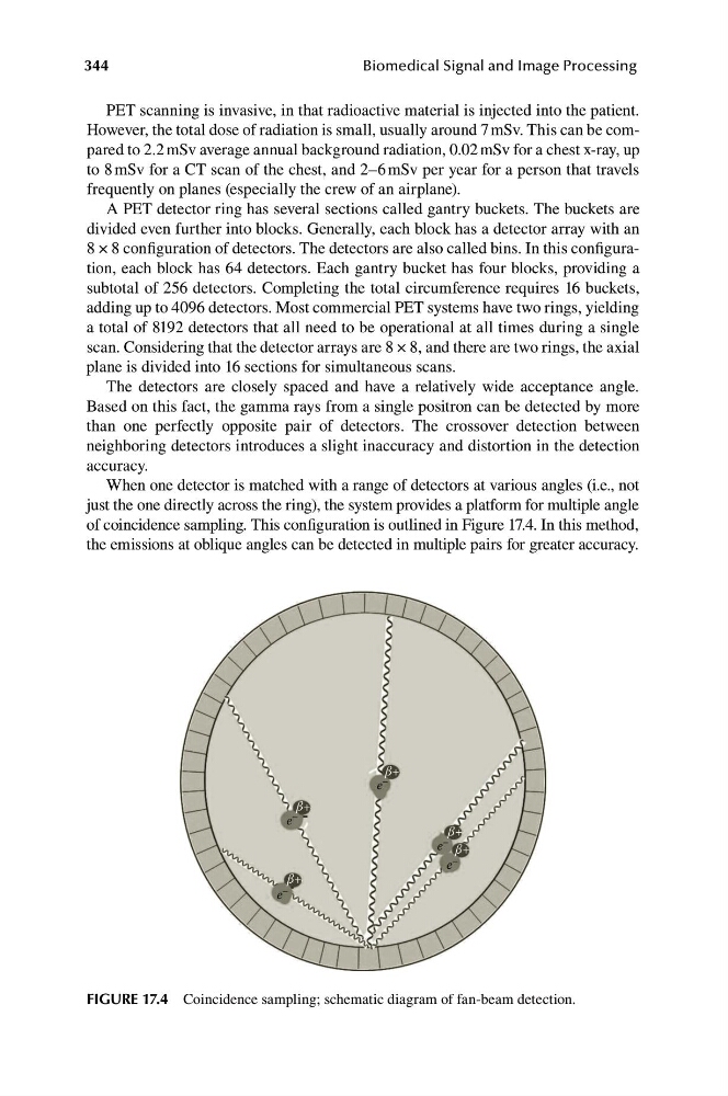
When one detector is matched with a range of detectors at various angles (i.e., not
just the one directly across the ring), the system provides a platform for multiple angle
of coincidence sampling. This configuration is outlined in Figure 17.4. In this method,
the emissions at oblique angles can be detected in multiple pairs for greater accuracy.
β+
β+
β+
β+
β+
e
–
e –
e –
e –
e –
FIGURE 17.4 Coincidence sampling; schematic diagram of fan-beam detection.
Biomedical Signal and Image Processing
PET scanning is invasive, in that radioactive material is injected into the patient.
However, the total dose of radiation is small, usually around 7 mSv. This can be compared to 2.2 mSv average annual background radiation, 0.02 mSv for a chest x-ray, up
to 8 mSv for a CT scan of the chest, and 2–6 mSv per year for a person that travels
frequently on planes (especially the crew of an airplane).
A PET detector ring has several sections called gantry buckets. The buckets are
divided even further into blocks. Generally, each block has a detector array with an
8 × 8 configuration of detectors. The detectors are also called bins. In this configuration, each block has 64 detectors. Each gantry bucket has four blocks, providing a
subtotal of 256 detectors. Completing the total circumference requires 16 buckets,
adding up to 4096 detectors. Most commercial PET systems have two rings, yielding
a total of 8192 detectors that all need to be operational at all times during a single
scan. Considering that the detector arrays are 8 × 8, and there are two rings, the axial
plane is divided into 16 sections for simultaneous scans.
The detectors are closely spaced and have a relatively wide acceptance angle.
Based on this fact, the gamma rays from a single positron can be detected by more
than one perfectly opposite pair of detectors. The crossover detection between
neighboring detectors introduces a slight inaccuracy and distortion in the detection
accuracy.
When one detector is matched with a range of detectors at various angles (i.e., not
just the one directly across the ring), the system provides a platform for multiple angle
of coincidence sampling. This configuration is outlined in Figure 17.4. In this method,
the emissions at oblique angles can be detected in multiple pairs for greater accuracy.
β+
β+
β+
β+
β+
e
–
e –
e –
e –
e –
FIGURE 17.4 Coincidence sampling; schematic diagram of fan-beam detection.