

328
Biomedical Signal and Image Processing
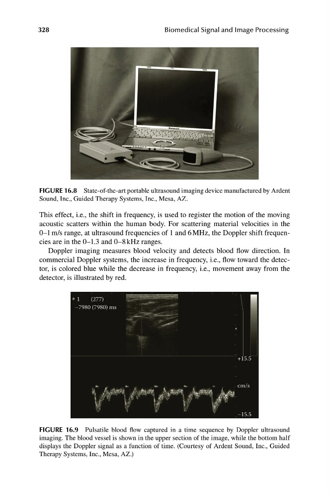
FIGURE 16.8 State-of-the-art portable ultrasound imaging device manufactured by Ardent
Sound, Inc., Guided Therapy Systems, Inc., Mesa, AZ.
This effect, i.e., the shift in frequency, is used to register the motion of the moving
acoustic scatters within the human body. For scattering material velocities in the
0–1 m/s range, at ultrasound frequencies of 1 and 6 MHz, the Doppler shift frequencies are in the 0–1.3 and 0–8 kHz ranges.
Doppler imaging measures blood velocity and detects blood flow direction. In
commercial Doppler systems, the increase in frequency, i.e., flow toward the detector, is colored blue while the decrease in frequency, i.e., movement away from the
detector, is illustrated by red.
1
(277)
–7980 (7980) ms
+15.5
cm/s
–15.5
FIGURE 16.9 Pulsatile blood flow captured in a time sequence by Doppler ultrasound
imaging. The blood vessel is shown in the upper section of the image, while the bottom half
displays the Doppler signal as a function of time. (Courtesy of Ardent Sound, Inc., Guided
Therapy Systems, Inc., Mesa, AZ.)
Biomedical Signal and Image Processing
FIGURE 16.8 State-of-the-art portable ultrasound imaging device manufactured by Ardent
Sound, Inc., Guided Therapy Systems, Inc., Mesa, AZ.
This effect, i.e., the shift in frequency, is used to register the motion of the moving
acoustic scatters within the human body. For scattering material velocities in the
0–1 m/s range, at ultrasound frequencies of 1 and 6 MHz, the Doppler shift frequencies are in the 0–1.3 and 0–8 kHz ranges.
Doppler imaging measures blood velocity and detects blood flow direction. In
commercial Doppler systems, the increase in frequency, i.e., flow toward the detector, is colored blue while the decrease in frequency, i.e., movement away from the
detector, is illustrated by red.
1
(277)
–7980 (7980) ms
+15.5
cm/s
–15.5
FIGURE 16.9 Pulsatile blood flow captured in a time sequence by Doppler ultrasound
imaging. The blood vessel is shown in the upper section of the image, while the bottom half
displays the Doppler signal as a function of time. (Courtesy of Ardent Sound, Inc., Guided
Therapy Systems, Inc., Mesa, AZ.)