

327
Ultrasound Imaging
RV
RV
LV
LV
C
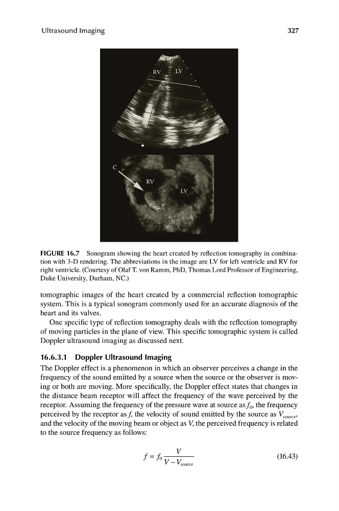
FIGURE 16.7 Sonogram showing the heart created by reflection tomography in combination with 3-D rendering. The abbreviations in the image are LV for left ventricle and RV for
right ventricle. (Courtesy of Olaf T. von Ramm, PhD, Thomas Lord Professor of Engineering,
Duke University, Durham, NC.)
tomographic images of the heart created by a commercial reflection tomographic
system. This is a typical sonogram commonly used for an accurate diagnosis of the
heart and its valves.
One specific type of reflection tomography deals with the reflection tomography
of moving particles in the plane of view. This specific tomographic system is called
Doppler ultrasound imaging as discussed next.
16.6.3.1 Doppler Ultrasound Imaging
The Doppler effect is a phenomenon in which an observer perceives a change in the
frequency of the sound emitted by a source when the source or the observer is moving or both are moving. More specifically, the Doppler effect states that changes in
the distance beam receptor will affect the frequency of the wave perceived by the
receptor. Assuming the frequency of the pressure wave at source as f 0 , the frequency
perceived by the receptor as f, the velocity of sound emitted by the source as V
,
source
and the velocity of the moving beam or object as V, the perceived frequency is related
to the source frequency as follows:
V
f = f
(16.43)
0
−
V V source
Ultrasound Imaging
RV
RV
LV
LV
C
FIGURE 16.7 Sonogram showing the heart created by reflection tomography in combination with 3-D rendering. The abbreviations in the image are LV for left ventricle and RV for
right ventricle. (Courtesy of Olaf T. von Ramm, PhD, Thomas Lord Professor of Engineering,
Duke University, Durham, NC.)
tomographic images of the heart created by a commercial reflection tomographic
system. This is a typical sonogram commonly used for an accurate diagnosis of the
heart and its valves.
One specific type of reflection tomography deals with the reflection tomography
of moving particles in the plane of view. This specific tomographic system is called
Doppler ultrasound imaging as discussed next.
16.6.3.1 Doppler Ultrasound Imaging
The Doppler effect is a phenomenon in which an observer perceives a change in the
frequency of the sound emitted by a source when the source or the observer is moving or both are moving. More specifically, the Doppler effect states that changes in
the distance beam receptor will affect the frequency of the wave perceived by the
receptor. Assuming the frequency of the pressure wave at source as f 0 , the frequency
perceived by the receptor as f, the velocity of sound emitted by the source as V
,
source
and the velocity of the moving beam or object as V, the perceived frequency is related
to the source frequency as follows:
V
f = f
(16.43)
0
−
V V source