

317
Ultrasound Imaging
and
P
2
q
t =
t
Z
=
A2 cos 1
(16.11)
P i Z A2 cos q 1 + Z A1 cos q 2
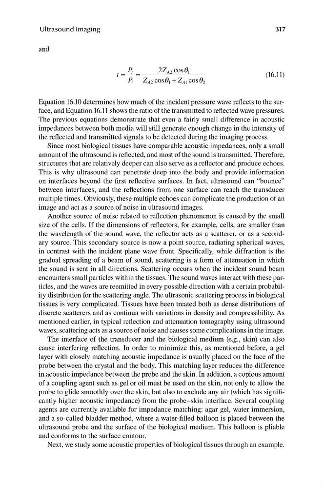
Equation 16.10 determines how much of the incident pressure wave reflects to the surface, and Equation 16.11 shows the ratio of the transmitted to reflected wave pressures.
The previous equations demonstrate that even a fairly small difference in acoustic
impedances between both media will still generate enough change in the intensity of
the reflected and transmitted signals to be detected during the imaging process.
Since most biological tissues have comparable acoustic impedances, only a small
amount of the ultrasound is reflected, and most of the sound is transmitted. Therefore,
structures that are relatively deeper can also serve as a reflector and produce echoes.
This is why ultrasound can penetrate deep into the body and provide information
on interfaces beyond the first reflective surfaces. In fact, ultrasound can “bounce”
between interfaces, and the reflections from one surface can reach the transducer
multiple times. Obviously, these multiple echoes can complicate the production of an
image and act as a source of noise in ultrasound images.
Another source of noise related to reflection phenomenon is caused by the small
size of the cells. If the dimensions of reflectors, for example, cells, are smaller than
the wavelength of the sound wave, the reflector acts as a scatterer, or as a secondary source. This secondary source is now a point source, radiating spherical waves,
in contrast with the incident plane wave front. Specifically, while diffraction is the
gradual spreading of a beam of sound, scattering is a form of attenuation in which
the sound is sent in all directions. Scattering occurs when the incident sound beam
encounters small particles within the tissues. The sound waves interact with these particles, and the waves are reemitted in every possible direction with a certain probability distribution for the scattering angle. The ultrasonic scattering process in biological
tissues is very complicated. Tissues have been treated both as dense distributions of
discrete scatterers and as continua with variations in density and compressibility. As
mentioned earlier, in typical reflection and attenuation tomography using ultrasound
waves, scattering acts as a source of noise and causes some complications in the image.
The interface of the transducer and the biological medium (e.g., skin) can also
cause interfering reflection. In order to minimize this, as mentioned before, a gel
layer with closely matching acoustic impedance is usually placed on the face of the
probe between the crystal and the body. This matching layer reduces the difference
in acoustic impedance between the probe and the skin. In addition, a copious amount
of a coupling agent such as gel or oil must be used on the skin, not only to allow the
probe to glide smoothly over the skin, but also to exclude any air (which has significantly higher acoustic impedance) from the probe–skin interface. Several coupling
agents are currently available for impedance matching: agar gel, water immersion,
and a so-called bladder method, where a water-filled balloon is placed between the
ultrasound probe and the surface of the biological medium. This balloon is pliable
and conforms to the surface contour.
Next, we study some acoustic properties of biological tissues through an example.
Ultrasound Imaging
and
P
2
q
t =
t
Z
=
A2 cos 1
(16.11)
P i Z A2 cos q 1 + Z A1 cos q 2
Equation 16.10 determines how much of the incident pressure wave reflects to the surface, and Equation 16.11 shows the ratio of the transmitted to reflected wave pressures.
The previous equations demonstrate that even a fairly small difference in acoustic
impedances between both media will still generate enough change in the intensity of
the reflected and transmitted signals to be detected during the imaging process.
Since most biological tissues have comparable acoustic impedances, only a small
amount of the ultrasound is reflected, and most of the sound is transmitted. Therefore,
structures that are relatively deeper can also serve as a reflector and produce echoes.
This is why ultrasound can penetrate deep into the body and provide information
on interfaces beyond the first reflective surfaces. In fact, ultrasound can “bounce”
between interfaces, and the reflections from one surface can reach the transducer
multiple times. Obviously, these multiple echoes can complicate the production of an
image and act as a source of noise in ultrasound images.
Another source of noise related to reflection phenomenon is caused by the small
size of the cells. If the dimensions of reflectors, for example, cells, are smaller than
the wavelength of the sound wave, the reflector acts as a scatterer, or as a secondary source. This secondary source is now a point source, radiating spherical waves,
in contrast with the incident plane wave front. Specifically, while diffraction is the
gradual spreading of a beam of sound, scattering is a form of attenuation in which
the sound is sent in all directions. Scattering occurs when the incident sound beam
encounters small particles within the tissues. The sound waves interact with these particles, and the waves are reemitted in every possible direction with a certain probability distribution for the scattering angle. The ultrasonic scattering process in biological
tissues is very complicated. Tissues have been treated both as dense distributions of
discrete scatterers and as continua with variations in density and compressibility. As
mentioned earlier, in typical reflection and attenuation tomography using ultrasound
waves, scattering acts as a source of noise and causes some complications in the image.
The interface of the transducer and the biological medium (e.g., skin) can also
cause interfering reflection. In order to minimize this, as mentioned before, a gel
layer with closely matching acoustic impedance is usually placed on the face of the
probe between the crystal and the body. This matching layer reduces the difference
in acoustic impedance between the probe and the skin. In addition, a copious amount
of a coupling agent such as gel or oil must be used on the skin, not only to allow the
probe to glide smoothly over the skin, but also to exclude any air (which has significantly higher acoustic impedance) from the probe–skin interface. Several coupling
agents are currently available for impedance matching: agar gel, water immersion,
and a so-called bladder method, where a water-filled balloon is placed between the
ultrasound probe and the surface of the biological medium. This balloon is pliable
and conforms to the surface contour.
Next, we study some acoustic properties of biological tissues through an example.