

318
Biomedical Signal and Image Processing
Example 16.1
Note the very large differences between the soft tissues and bone and air.
Assuming that the incident wave is perpendicular to the surface of the interface,
we use the reflection equation to compare the reflection between two types of
soft tissue, i.e., fat and muscle, with the reflection between a soft tissue and a hard
tissue, i.e., brain to skull.
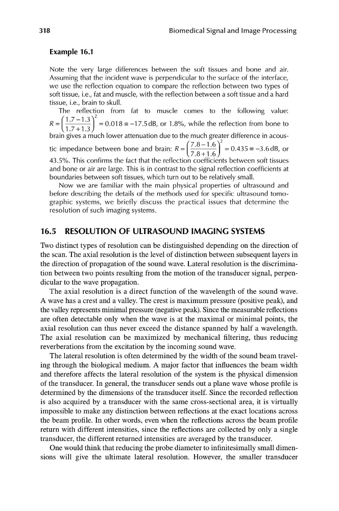
The reflection from fat to muscle comes to the following value:
1 3⎞
2
⎛ 1 7
. − .
R =
= 0 018
.
≡− 17 5 d B , or 1.8%, while the reflection from bone to
.
⎝ ⎜
⎠ ⎟
1 7
. + 1 3
.
brain gives a much lower attenuation due to the much greater difference in acous1 6⎞
2
⎛ 7 8
. − .
.
tic impedance between bone and brain: R =
= 0 435
.
≡− 3 6 d B , or
⎝ ⎜ 7 8
. + 1 6⎠
⎟
.
43.5%. This confirms the fact that the reflection coefficients between soft tissues
and bone or air are large. This is in contrast to the signal reflection coefficients at
boundaries between soft tissues, which turn out to be relatively small.
Now we are familiar with the main physical properties of ultrasound and
before describing the details of the methods used for specific ultrasound tomographic systems, we briefly discuss the practical issues that determine the
resolution of such imaging systems.
16.5 RESOLUTION OF ULTRASOUND IMAGING SYSTEMS
Two distinct types of resolution can be distinguished depending on the direction of
the scan. The axial resolution is the level of distinction between subsequent layers in
the direction of propagation of the sound wave. Lateral resolution is the discrimination between two points resulting from the motion of the transducer signal, perpendicular to the wave propagation.
The axial resolution is a direct function of the wavelength of the sound wave.
A wave has a crest and a valley. The crest is maximum pressure (positive peak), and
the valley represents minimal pressure (negative peak). Since the measurable reflections
are often detectable only when the wave is at the maximal or minimal points, the
axial resolution can thus never exceed the distance spanned by half a wavelength.
The axial resolution can be maximized by mechanical filtering, thus reducing
reverberations from the excitation by the incoming sound wave.
The lateral resolution is often determined by the width of the sound beam traveling through the biological medium. A major factor that influences the beam width
and therefore affects the lateral resolution of the system is the physical dimension
of the transducer. In general, the transducer sends out a plane wave whose profile is
determined by the dimensions of the transducer itself. Since the recorded reflection
is also acquired by a transducer with the same cross-sectional area, it is virtually
impossible to make any distinction between reflections at the exact locations across
the beam profile. In other words, even when the reflections across the beam profile
return with different intensities, since the reflections are collected by only a single
transducer, the different returned intensities are averaged by the transducer.
One would think that reducing the probe diameter to infinitesimally small dimensions will give the ultimate lateral resolution. However, the smaller transducer