

m
m
m
m
m
m
m
m
286
Biomedical Signal and Image Processing
of each other. However, if the human body is exposed to an external magnetic field,
these ensembles start showing measurable magnetic effects.
In NMR imaging, all nuclei with an odd number of protons are immersed in a
static magnetic field, H 0 , in the z-direction. In MRI technology, a very strong constant magnetic field with a typical magnitude between 0.2 and about 3 T is used
as H 0 . Knowing that the earth’s magnetic field is approximately 5 × 10 −5 T, it can
be seen that the applied magnetic field is very strong. The presence of this strong
external magnetic field creates a net magnetic field in the microscopic elements. The
resultant nuclear magnetization, M, is the total magnetic moment of all the microscopic units, e.g., elements with an odd number of charged particles in the nuclei.
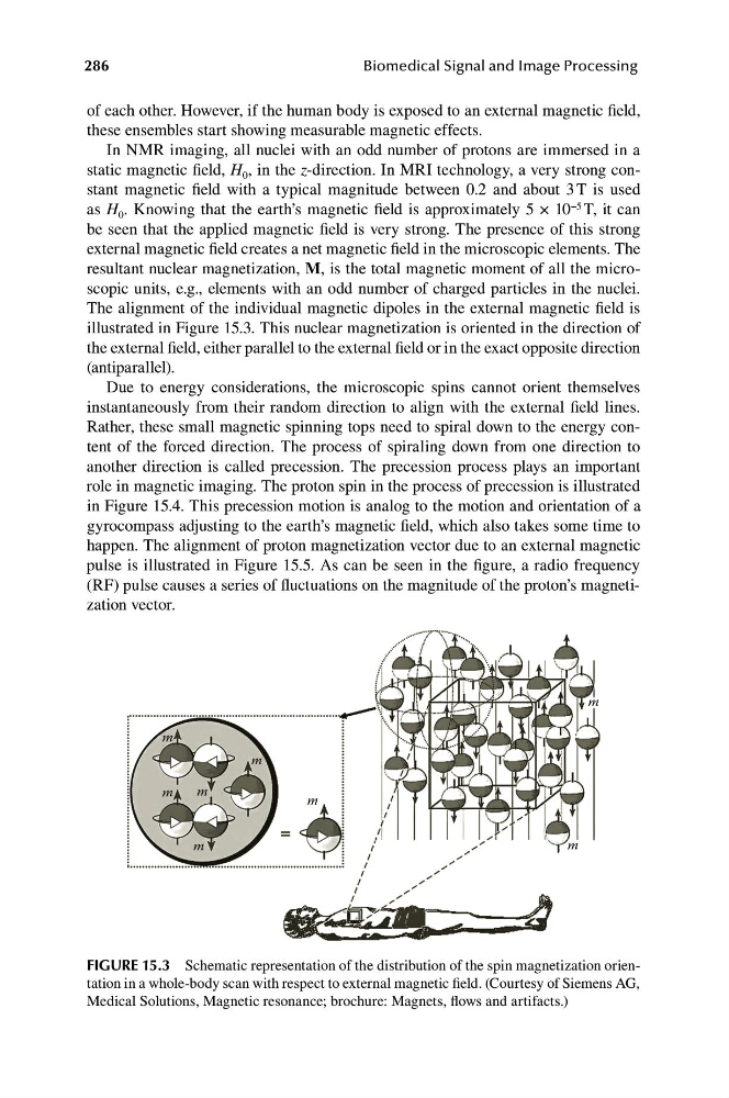
The alignment of the individual magnetic dipoles in the external magnetic field is
illustrated in Figure 15.3. This nuclear magnetization is oriented in the direction of
the external field, either parallel to the external field or in the exact opposite direction
(antiparallel).
Due to energy considerations, the microscopic spins cannot orient themselves
instantaneously from their random direction to align with the external field lines.
Rather, these small magnetic spinning tops need to spiral down to the energy content of the forced direction. The process of spiraling down from one direction to
another direction is called precession. The precession process plays an important
role in magnetic imaging. The proton spin in the process of precession is illustrated
in Figure 15.4. This precession motion is analog to the motion and orientation of a
gyrocompass adjusting to the earth’s magnetic field, which also takes some time to
happen. The alignment of proton magnetization vector due to an external magnetic
pulse is illustrated in Figure 15.5. As can be seen in the figure, a radio frequency
(RF) pulse causes a series of fluctuations on the magnitude of the proton’s magnetization vector.
FIGURE 15.3 Schematic representation of the distribution of the spin magnetization orientation in a whole-body scan with respect to external magnetic field. (Courtesy of Siemens AG,
Medical Solutions, Magnetic resonance; brochure: Magnets, flows and artifacts.)
m
m
m
m
m
m
m
286
Biomedical Signal and Image Processing
of each other. However, if the human body is exposed to an external magnetic field,
these ensembles start showing measurable magnetic effects.
In NMR imaging, all nuclei with an odd number of protons are immersed in a
static magnetic field, H 0 , in the z-direction. In MRI technology, a very strong constant magnetic field with a typical magnitude between 0.2 and about 3 T is used
as H 0 . Knowing that the earth’s magnetic field is approximately 5 × 10 −5 T, it can
be seen that the applied magnetic field is very strong. The presence of this strong
external magnetic field creates a net magnetic field in the microscopic elements. The
resultant nuclear magnetization, M, is the total magnetic moment of all the microscopic units, e.g., elements with an odd number of charged particles in the nuclei.
The alignment of the individual magnetic dipoles in the external magnetic field is
illustrated in Figure 15.3. This nuclear magnetization is oriented in the direction of
the external field, either parallel to the external field or in the exact opposite direction
(antiparallel).
Due to energy considerations, the microscopic spins cannot orient themselves
instantaneously from their random direction to align with the external field lines.
Rather, these small magnetic spinning tops need to spiral down to the energy content of the forced direction. The process of spiraling down from one direction to
another direction is called precession. The precession process plays an important
role in magnetic imaging. The proton spin in the process of precession is illustrated
in Figure 15.4. This precession motion is analog to the motion and orientation of a
gyrocompass adjusting to the earth’s magnetic field, which also takes some time to
happen. The alignment of proton magnetization vector due to an external magnetic
pulse is illustrated in Figure 15.5. As can be seen in the figure, a radio frequency
(RF) pulse causes a series of fluctuations on the magnitude of the proton’s magnetization vector.
FIGURE 15.3 Schematic representation of the distribution of the spin magnetization orientation in a whole-body scan with respect to external magnetic field. (Courtesy of Siemens AG,
Medical Solutions, Magnetic resonance; brochure: Magnets, flows and artifacts.)