

273
X-Ray Imaging and Computed Tomography
In addition, unlike the conventional x-ray where in order to form an image, only a single
x-ray scan from only a single angle is conducted, in CT, several scans from many angles
are taken to form a single image. This process was described in Chapter 13.
CT scanning exposes the patient to moderate to high levels of radiation, mostly
due to the extended exposure time. In CT, a repetitive procedure is followed that
scans the biological tissues from many directions by revolving both the source and
the scintillation counter detectors around the body in a circular motion.
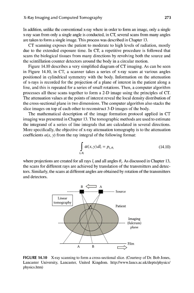
Figure 14.10 describes a very simplified diagram of CT imaging. As can be seen
in Figure 14.10, in CT, a scanner takes a series of x-ray scans at various angles
positioned in cylindrical symmetry with the body. Information on the attenuation
of x-rays is recorded for the projection of a plane of interest in the patient along a
line, and this is repeated for a series of small rotations. Then, a computer algorithm
processes all these scans together to form a 2-D image using the principles of CT.
The attenuation values at the points of interest reveal the local density distribution of
the cross-sectional plane in two dimensions. The computer algorithm also stacks the
slice images on top of each other to reconstruct 3-D images of the body.
The mathematical description of the image formation protocol applied in CT
imaging was presented in Chapter 13. The tomographic methods are used to estimate
the integrand of a series of line integrals that are calculated in several directions.
More specifically, the objective of x-ray attenuation tomography is to the attenuation
coefficients α(x, y) from the ray integral of the following format:
∫
a( , ) = p l i q i
x y dl i
,
(14.10)
l ,q
i i
where projections are created for all rays l i and all angles θ i . As discussed in Chapter 13,
the scans for different rays are achieved by translation of the transmitters and detectors. Similarly, the scans at different angles are obtained by rotation of the transmitters
and detectors.
Linear
tomography
B
A
Source
Patient
Imaging
(fulcrum)
plane
Film
A
B
FIGURE 14.10 X-ray scanning to form a cross-sectional slice. (Courtesy of Dr. Bob Jones,
Lancaster University, Lancaster, United Kingdom. http://www.lancs.ac.uk/depts/physics/
physics.htm)
X-Ray Imaging and Computed Tomography
In addition, unlike the conventional x-ray where in order to form an image, only a single
x-ray scan from only a single angle is conducted, in CT, several scans from many angles
are taken to form a single image. This process was described in Chapter 13.
CT scanning exposes the patient to moderate to high levels of radiation, mostly
due to the extended exposure time. In CT, a repetitive procedure is followed that
scans the biological tissues from many directions by revolving both the source and
the scintillation counter detectors around the body in a circular motion.
Figure 14.10 describes a very simplified diagram of CT imaging. As can be seen
in Figure 14.10, in CT, a scanner takes a series of x-ray scans at various angles
positioned in cylindrical symmetry with the body. Information on the attenuation
of x-rays is recorded for the projection of a plane of interest in the patient along a
line, and this is repeated for a series of small rotations. Then, a computer algorithm
processes all these scans together to form a 2-D image using the principles of CT.
The attenuation values at the points of interest reveal the local density distribution of
the cross-sectional plane in two dimensions. The computer algorithm also stacks the
slice images on top of each other to reconstruct 3-D images of the body.
The mathematical description of the image formation protocol applied in CT
imaging was presented in Chapter 13. The tomographic methods are used to estimate
the integrand of a series of line integrals that are calculated in several directions.
More specifically, the objective of x-ray attenuation tomography is to the attenuation
coefficients α(x, y) from the ray integral of the following format:
∫
a( , ) = p l i q i
x y dl i
,
(14.10)
l ,q
i i
where projections are created for all rays l i and all angles θ i . As discussed in Chapter 13,
the scans for different rays are achieved by translation of the transmitters and detectors. Similarly, the scans at different angles are obtained by rotation of the transmitters
and detectors.
Linear
tomography
B
A
Source
Patient
Imaging
(fulcrum)
plane
Film
A
B
FIGURE 14.10 X-ray scanning to form a cross-sectional slice. (Courtesy of Dr. Bob Jones,
Lancaster University, Lancaster, United Kingdom. http://www.lancs.ac.uk/depts/physics/
physics.htm)