

d
Biological
medium
X-ray film
Area of anatomic
of interest
Source
L
l
p
272
Biomedical Signal and Image Processing
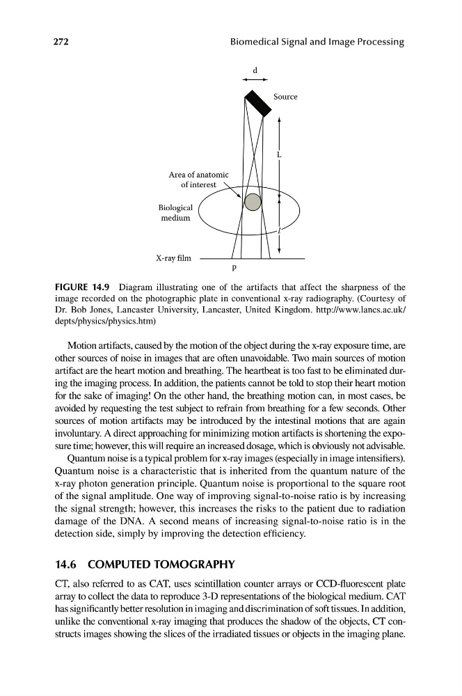
FIGURE 14.9 Diagram illustrating one of the artifacts that affect the sharpness of the
image recorded on the photographic plate in conventional x-ray radiography. (Courtesy of
Dr. Bob Jones, Lancaster University, Lancaster, United Kingdom. http://www.lancs.ac.uk/
depts/physics/physics.htm)
Motion artifacts, caused by the motion of the object during the x-ray exposure time, are
other sources of noise in images that are often unavoidable. Two main sources of motion
artifact are the heart motion and breathing. The heartbeat is too fast to be eliminated during the imaging process. In addition, the patients cannot be told to stop their heart motion
for the sake of imaging! On the other hand, the breathing motion can, in most cases, be
avoided by requesting the test subject to refrain from breathing for a few seconds. Other
sources of motion artifacts may be introduced by the intestinal motions that are again
involuntary. A direct approaching for minimizing motion artifacts is shortening the exposure time; however, this will require an increased dosage, which is obviously not advisable.
Quantum noise is a typical problem for x-ray images (especially in image intensifiers).
Quantum noise is a characteristic that is inherited from the quantum nature of the
x-ray photon generation principle. Quantum noise is proportional to the square root
of the signal amplitude. One way of improving signal-to-noise ratio is by increasing
the signal strength; however, this increases the risks to the patient due to radiation
damage of the DNA. A second means of increasing signal-to-noise ratio is in the
detection side, simply by improving the detection efficiency.
14.6 COMPUTED TOMOGRAPHY
CT, also referred to as CAT, uses scintillation counter arrays or CCD-fluorescent plate
array to collect the data to reproduce 3-D representations of the biological medium. CAT
has significantly better resolution in imaging and discrimination of soft tissues. In addition,
unlike the conventional x-ray imaging that produces the shadow of the objects, CT constructs images showing the slices of the irradiated tissues or objects in the imaging plane.
Biological
medium
X-ray film
Area of anatomic
of interest
Source
L
l
p
272
Biomedical Signal and Image Processing
FIGURE 14.9 Diagram illustrating one of the artifacts that affect the sharpness of the
image recorded on the photographic plate in conventional x-ray radiography. (Courtesy of
Dr. Bob Jones, Lancaster University, Lancaster, United Kingdom. http://www.lancs.ac.uk/
depts/physics/physics.htm)
Motion artifacts, caused by the motion of the object during the x-ray exposure time, are
other sources of noise in images that are often unavoidable. Two main sources of motion
artifact are the heart motion and breathing. The heartbeat is too fast to be eliminated during the imaging process. In addition, the patients cannot be told to stop their heart motion
for the sake of imaging! On the other hand, the breathing motion can, in most cases, be
avoided by requesting the test subject to refrain from breathing for a few seconds. Other
sources of motion artifacts may be introduced by the intestinal motions that are again
involuntary. A direct approaching for minimizing motion artifacts is shortening the exposure time; however, this will require an increased dosage, which is obviously not advisable.
Quantum noise is a typical problem for x-ray images (especially in image intensifiers).
Quantum noise is a characteristic that is inherited from the quantum nature of the
x-ray photon generation principle. Quantum noise is proportional to the square root
of the signal amplitude. One way of improving signal-to-noise ratio is by increasing
the signal strength; however, this increases the risks to the patient due to radiation
damage of the DNA. A second means of increasing signal-to-noise ratio is in the
detection side, simply by improving the detection efficiency.
14.6 COMPUTED TOMOGRAPHY
CT, also referred to as CAT, uses scintillation counter arrays or CCD-fluorescent plate
array to collect the data to reproduce 3-D representations of the biological medium. CAT
has significantly better resolution in imaging and discrimination of soft tissues. In addition,
unlike the conventional x-ray imaging that produces the shadow of the objects, CT constructs images showing the slices of the irradiated tissues or objects in the imaging plane.