

+ –
Differential
amplifier
– +
Differential
amplifier
239
Other Biomedical Signals
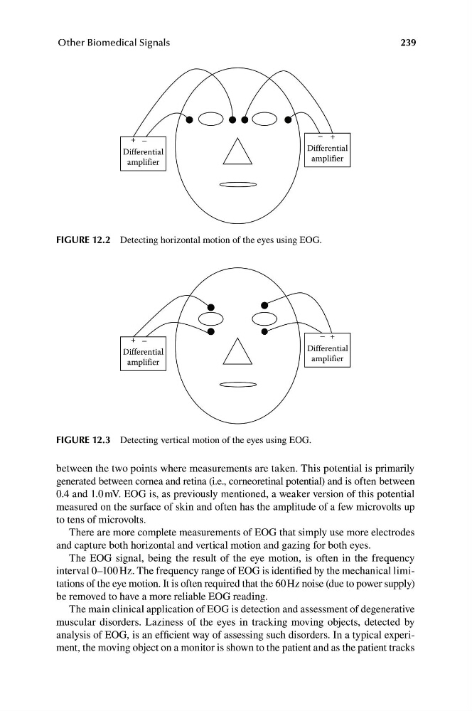
FIGURE 12.2 Detecting horizontal motion of the eyes using EOG.
– +
+ –
Differential
Differential
amplifier
amplifier
FIGURE 12.3 Detecting vertical motion of the eyes using EOG.
between the two points where measurements are taken. This potential is primarily
generated between cornea and retina (i.e., corneoretinal potential) and is often between
0.4 and 1.0 mV. EOG is, as previously mentioned, a weaker version of this potential
measured on the surface of skin and often has the amplitude of a few microvolts up
to tens of microvolts.
There are more complete measurements of EOG that simply use more electrodes
and capture both horizontal and vertical motion and gazing for both eyes.
The EOG signal, being the result of the eye motion, is often in the frequency
interval 0–100 Hz. The frequency range of EOG is identified by the mechanical limitations of the eye motion. It is often required that the 60 Hz noise (due to power supply)
be removed to have a more reliable EOG reading.
The main clinical application of EOG is detection and assessment of degenerative
muscular disorders. Laziness of the eyes in tracking moving objects, detected by
analysis of EOG, is an efficient way of assessing such disorders. In a typical experiment, the moving object on a monitor is shown to the patient and as the patient tracks
Differential
amplifier
– +
Differential
amplifier
239
Other Biomedical Signals
FIGURE 12.2 Detecting horizontal motion of the eyes using EOG.
– +
+ –
Differential
Differential
amplifier
amplifier
FIGURE 12.3 Detecting vertical motion of the eyes using EOG.
between the two points where measurements are taken. This potential is primarily
generated between cornea and retina (i.e., corneoretinal potential) and is often between
0.4 and 1.0 mV. EOG is, as previously mentioned, a weaker version of this potential
measured on the surface of skin and often has the amplitude of a few microvolts up
to tens of microvolts.
There are more complete measurements of EOG that simply use more electrodes
and capture both horizontal and vertical motion and gazing for both eyes.
The EOG signal, being the result of the eye motion, is often in the frequency
interval 0–100 Hz. The frequency range of EOG is identified by the mechanical limitations of the eye motion. It is often required that the 60 Hz noise (due to power supply)
be removed to have a more reliable EOG reading.
The main clinical application of EOG is detection and assessment of degenerative
muscular disorders. Laziness of the eyes in tracking moving objects, detected by
analysis of EOG, is an efficient way of assessing such disorders. In a typical experiment, the moving object on a monitor is shown to the patient and as the patient tracks