

238
Biomedical Signal and Image Processing
Pressure
Time
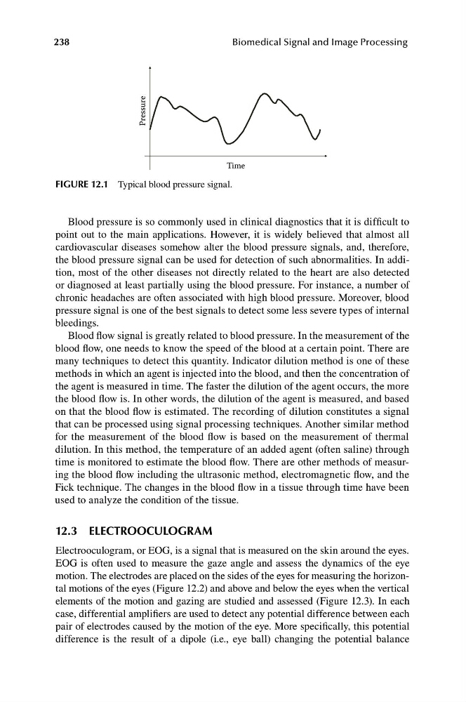
FIGURE 12.1 Typical blood pressure signal.
Blood pressure is so commonly used in clinical diagnostics that it is difficult to
point out to the main applications. However, it is widely believed that almost all
cardiovascular diseases somehow alter the blood pressure signals, and, therefore,
the blood pressure signal can be used for detection of such abnormalities. In addition, most of the other diseases not directly related to the heart are also detected
or diagnosed at least partially using the blood pressure. For instance, a number of
chronic headaches are often associated with high blood pressure. Moreover, blood
pressure signal is one of the best signals to detect some less severe types of internal
bleedings.
Blood flow signal is greatly related to blood pressure. In the measurement of the
blood flow, one needs to know the speed of the blood at a certain point. There are
many techniques to detect this quantity. Indicator dilution method is one of these
methods in which an agent is injected into the blood, and then the concentration of
the agent is measured in time. The faster the dilution of the agent occurs, the more
the blood flow is. In other words, the dilution of the agent is measured, and based
on that the blood flow is estimated. The recording of dilution constitutes a signal
that can be processed using signal processing techniques. Another similar method
for the measurement of the blood flow is based on the measurement of thermal
dilution. In this method, the temperature of an added agent (often saline) through
time is monitored to estimate the blood flow. There are other methods of measuring the blood flow including the ultrasonic method, electromagnetic flow, and the
Fick technique. The changes in the blood flow in a tissue through time have been
used to analyze the condition of the tissue.
12.3 ELECTROOCULOGRAM
Electrooculogram, or EOG, is a signal that is measured on the skin around the eyes.
EOG is often used to measure the gaze angle and assess the dynamics of the eye
motion. The electrodes are placed on the sides of the eyes for measuring the horizontal motions of the eyes (Figure 12.2) and above and below the eyes when the vertical
elements of the motion and gazing are studied and assessed (Figure 12.3). In each
case, differential amplifiers are used to detect any potential difference between each
pair of electrodes caused by the motion of the eye. More specifically, this potential
difference is the result of a dipole (i.e., eye ball) changing the potential balance
Biomedical Signal and Image Processing
Pressure
Time
FIGURE 12.1 Typical blood pressure signal.
Blood pressure is so commonly used in clinical diagnostics that it is difficult to
point out to the main applications. However, it is widely believed that almost all
cardiovascular diseases somehow alter the blood pressure signals, and, therefore,
the blood pressure signal can be used for detection of such abnormalities. In addition, most of the other diseases not directly related to the heart are also detected
or diagnosed at least partially using the blood pressure. For instance, a number of
chronic headaches are often associated with high blood pressure. Moreover, blood
pressure signal is one of the best signals to detect some less severe types of internal
bleedings.
Blood flow signal is greatly related to blood pressure. In the measurement of the
blood flow, one needs to know the speed of the blood at a certain point. There are
many techniques to detect this quantity. Indicator dilution method is one of these
methods in which an agent is injected into the blood, and then the concentration of
the agent is measured in time. The faster the dilution of the agent occurs, the more
the blood flow is. In other words, the dilution of the agent is measured, and based
on that the blood flow is estimated. The recording of dilution constitutes a signal
that can be processed using signal processing techniques. Another similar method
for the measurement of the blood flow is based on the measurement of thermal
dilution. In this method, the temperature of an added agent (often saline) through
time is monitored to estimate the blood flow. There are other methods of measuring the blood flow including the ultrasonic method, electromagnetic flow, and the
Fick technique. The changes in the blood flow in a tissue through time have been
used to analyze the condition of the tissue.
12.3 ELECTROOCULOGRAM
Electrooculogram, or EOG, is a signal that is measured on the skin around the eyes.
EOG is often used to measure the gaze angle and assess the dynamics of the eye
motion. The electrodes are placed on the sides of the eyes for measuring the horizontal motions of the eyes (Figure 12.2) and above and below the eyes when the vertical
elements of the motion and gazing are studied and assessed (Figure 12.3). In each
case, differential amplifiers are used to detect any potential difference between each
pair of electrodes caused by the motion of the eye. More specifically, this potential
difference is the result of a dipole (i.e., eye ball) changing the potential balance