

201
Electroencephalogram
10.3.1 EEG FREQUENCY SPECTRUM
The EEG signal is often interpreted based on the presence and absence of particular
waves with known frequencies. The presence or absence of these waves indicates
certain physiological conditions that are extremely useful in diagnostics. The typical waves classified in the EEG signal are alpha (α), beta (β), delta (δ), and theta (Θ)
waves. The presence and strength of each of these waves can be easily detected
using discrete Fourier transform (DFT) of the EEG signal (in deterministic analysis
of the signal) or using the power spectra of the EEG signal (in stochastic processing
of EEG). The definition of each wave identifies the formation of band-pass filters
needed to extract these waves.
The normal adult, in a physically relaxed state while having the eyes closed, produces a rhythmical brain wave with a frequency of 8–14 Hz, which is called alpha
wave. Generally, the alpha wave disappears when the eyes are opened; however, they
may still be present if the person is in a state of extreme relaxation. Since these waves
were discovered first, they were labeled as alpha waves. From the definition of the
alpha wave, it is clear that in order to extract this wave component, one needs to pass
the EEG through a band-pass filter with the pass band set to 8–14 Hz.
The frequency band of the beta waves is 14–50 Hz. The beta waves are prevalent
in the regions of parietal and frontal lobes. Beta waves are often associated with
problem solving and logical thinking. As will be discussed later, beta wave is the
major component observed in an important stage of healthy sleep. In order to extract
the beta wave, a band-pass filter with the pass band set to 14–50 Hz must be used.
The delta waves have a frequency range of 0.5–4 Hz and are detected in infants
and sleeping adults. The band-pass filter needed to extract delta wave has a pass band
of 0.5–4 Hz.
The theta waves fall in the frequency range of 4–8 Hz and are measured in resting
and sleeping adults and in children while awake. This wave can be separated using
a 4–8 Hz band-pass filter.
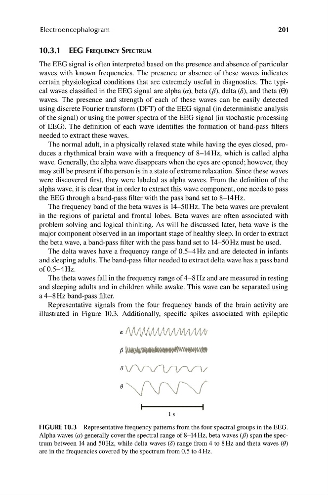
Representative signals from the four frequency bands of the brain activity are
illustrated in Figure 10.3. Additionally, specific spikes associated with epileptic
α
β
δ
θ
1 s
FIGURE 10.3 Representative frequency patterns from the four spectral groups in the EEG.
Alpha waves (α) generally cover the spectral range of 8–14 Hz, beta waves (β) span the spectrum between 14 and 50 Hz, while delta waves (δ) range from 4 to 8 Hz and theta waves (θ)
are in the frequencies covered by the spectrum from 0.5 to 4 Hz.
Electroencephalogram
10.3.1 EEG FREQUENCY SPECTRUM
The EEG signal is often interpreted based on the presence and absence of particular
waves with known frequencies. The presence or absence of these waves indicates
certain physiological conditions that are extremely useful in diagnostics. The typical waves classified in the EEG signal are alpha (α), beta (β), delta (δ), and theta (Θ)
waves. The presence and strength of each of these waves can be easily detected
using discrete Fourier transform (DFT) of the EEG signal (in deterministic analysis
of the signal) or using the power spectra of the EEG signal (in stochastic processing
of EEG). The definition of each wave identifies the formation of band-pass filters
needed to extract these waves.
The normal adult, in a physically relaxed state while having the eyes closed, produces a rhythmical brain wave with a frequency of 8–14 Hz, which is called alpha
wave. Generally, the alpha wave disappears when the eyes are opened; however, they
may still be present if the person is in a state of extreme relaxation. Since these waves
were discovered first, they were labeled as alpha waves. From the definition of the
alpha wave, it is clear that in order to extract this wave component, one needs to pass
the EEG through a band-pass filter with the pass band set to 8–14 Hz.
The frequency band of the beta waves is 14–50 Hz. The beta waves are prevalent
in the regions of parietal and frontal lobes. Beta waves are often associated with
problem solving and logical thinking. As will be discussed later, beta wave is the
major component observed in an important stage of healthy sleep. In order to extract
the beta wave, a band-pass filter with the pass band set to 14–50 Hz must be used.
The delta waves have a frequency range of 0.5–4 Hz and are detected in infants
and sleeping adults. The band-pass filter needed to extract delta wave has a pass band
of 0.5–4 Hz.
The theta waves fall in the frequency range of 4–8 Hz and are measured in resting
and sleeping adults and in children while awake. This wave can be separated using
a 4–8 Hz band-pass filter.
Representative signals from the four frequency bands of the brain activity are
illustrated in Figure 10.3. Additionally, specific spikes associated with epileptic
α
β
δ
θ
1 s
FIGURE 10.3 Representative frequency patterns from the four spectral groups in the EEG.
Alpha waves (α) generally cover the spectral range of 8–14 Hz, beta waves (β) span the spectrum between 14 and 50 Hz, while delta waves (δ) range from 4 to 8 Hz and theta waves (θ)
are in the frequencies covered by the spectrum from 0.5 to 4 Hz.