

189
Electrocardiogram
I
aV R
V1
V4
II
aV L
V2
V5
III
aV F
V3
V6
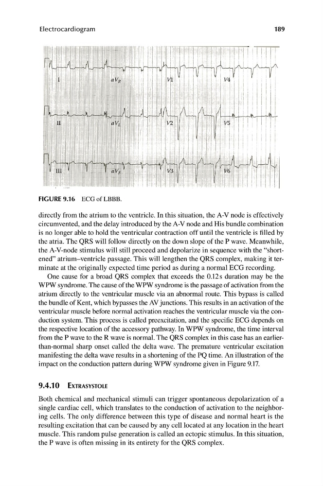
FIGURE 9.16 ECG of LBBB.
directly from the atrium to the ventricle. In this situation, the A-V node is effectively
circumvented, and the delay introduced by the A-V node and His bundle combination
is no longer able to hold the ventricular contraction off until the ventricle is filled by
the atria. The QRS will follow directly on the down slope of the P wave. Meanwhile,
the A-V-node stimulus will still proceed and depolarize in sequence with the “shortened” atrium–ventricle passage. This will lengthen the QRS complex, making it terminate at the originally expected time period as during a normal ECG recording.
One cause for a broad QRS complex that exceeds the 0.12s duration may be the
WPW syndrome. The cause of the WPW syndrome is the passage of activation from the
atrium directly to the ventricular muscle via an abnormal route. This bypass is called
the bundle of Kent, which bypasses the AV junctions. This results in an activation of the
ventricular muscle before normal activation reaches the ventricular muscle via the conduction system. This process is called preexcitation, and the specific ECG depends on
the respective location of the accessory pathway. In WPW syndrome, the time interval
from the P wave to the R wave is normal. The QRS complex in this case has an earlierthan-normal sharp onset called the delta wave. The premature ventricular excitation
manifesting the delta wave results in a shortening of the PQ time. An illustration of the
impact on the conduction pattern during WPW syndrome given in Figure 9.17.
9.4.10 EXTRASYSTOLE
Both chemical and mechanical stimuli can trigger spontaneous depolarization of a
single cardiac cell, which translates to the conduction of activation to the neighboring cells. The only difference between this type of disease and normal heart is the
resulting excitation that can be caused by any cell located at any location in the heart
muscle. This random pulse generation is called an ectopic stimulus. In this situation,
the P wave is often missing in its entirety for the QRS complex.
Electrocardiogram
I
aV R
V1
V4
II
aV L
V2
V5
III
aV F
V3
V6
FIGURE 9.16 ECG of LBBB.
directly from the atrium to the ventricle. In this situation, the A-V node is effectively
circumvented, and the delay introduced by the A-V node and His bundle combination
is no longer able to hold the ventricular contraction off until the ventricle is filled by
the atria. The QRS will follow directly on the down slope of the P wave. Meanwhile,
the A-V-node stimulus will still proceed and depolarize in sequence with the “shortened” atrium–ventricle passage. This will lengthen the QRS complex, making it terminate at the originally expected time period as during a normal ECG recording.
One cause for a broad QRS complex that exceeds the 0.12s duration may be the
WPW syndrome. The cause of the WPW syndrome is the passage of activation from the
atrium directly to the ventricular muscle via an abnormal route. This bypass is called
the bundle of Kent, which bypasses the AV junctions. This results in an activation of the
ventricular muscle before normal activation reaches the ventricular muscle via the conduction system. This process is called preexcitation, and the specific ECG depends on
the respective location of the accessory pathway. In WPW syndrome, the time interval
from the P wave to the R wave is normal. The QRS complex in this case has an earlierthan-normal sharp onset called the delta wave. The premature ventricular excitation
manifesting the delta wave results in a shortening of the PQ time. An illustration of the
impact on the conduction pattern during WPW syndrome given in Figure 9.17.
9.4.10 EXTRASYSTOLE
Both chemical and mechanical stimuli can trigger spontaneous depolarization of a
single cardiac cell, which translates to the conduction of activation to the neighboring cells. The only difference between this type of disease and normal heart is the
resulting excitation that can be caused by any cell located at any location in the heart
muscle. This random pulse generation is called an ectopic stimulus. In this situation,
the P wave is often missing in its entirety for the QRS complex.