

188
Biomedical Signal and Image Processing
I
V1
V4
aV R
II
V2
V5
aV L
III
aV F
V3
V6
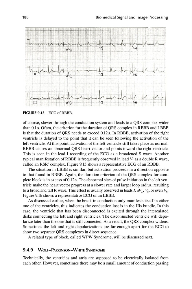
FIGURE 9.15 ECG of RBBB.
of course, slower through the conduction system and leads to a QRS complex wider
than 0.1 s. Often, the criterion for the duration of QRS complex in RBBB and LBBB
is that the duration of QRS needs to exceed 0.12 s. In RBBB, activation of the right
ventricle is delayed to the point that it can be seen following the activation of the
left ventricle. At this point, activation of the left ventricle still takes place as normal.
RBBB causes an abnormal QRS heart vector and points toward the right ventricle.
This is seen in the lead I recording of the ECG as a broadened S wave. Another
typical manifestation of RBBB is frequently observed in lead V 1 as a double R wave,
called an RSR’ complex. Figure 9.15 shows a representative ECG of an RBBB.
The situation in LBBB is similar, but activation proceeds in a direction opposite
to that found in RBBB. Again, the duration criterion of the QRS complex for complete block is in excess of 0.12 s. The abnormal sites of pulse initiation in the left ventricle make the heart vector progress at a slower rate and larger loop radius, resulting
to a broad and tall R wave. This effect is usually observed in leads I, aV L , V 5 , or even V 6 .
Figure 9.16 shows a representative ECG of an LBBB.
As discussed earlier, when the break in conduction only manifests itself in either
one of the ventricles, this indicates the conduction lost is in the His bundle. In this
case, the ventricle that has been disconnected is excited through the intercalated
disks connecting the left and right ventricles. The disconnected ventricle will depolarize later than the one that is still connected. As a result, the QRS complex widens.
Sometimes the left and right depolarizations are far enough apart for the ECG to
show two separate QRS complexes in direct sequence.
A related type of block, called WPW Syndrome, will be discussed next.
9.4.9 WOLF–PARKINSON–WHITE SYNDROME
Technically, the ventricles and atria are supposed to be electrically isolated from
each other. However, sometimes there may be a small amount of conduction passing
Biomedical Signal and Image Processing
I
V1
V4
aV R
II
V2
V5
aV L
III
aV F
V3
V6
FIGURE 9.15 ECG of RBBB.
of course, slower through the conduction system and leads to a QRS complex wider
than 0.1 s. Often, the criterion for the duration of QRS complex in RBBB and LBBB
is that the duration of QRS needs to exceed 0.12 s. In RBBB, activation of the right
ventricle is delayed to the point that it can be seen following the activation of the
left ventricle. At this point, activation of the left ventricle still takes place as normal.
RBBB causes an abnormal QRS heart vector and points toward the right ventricle.
This is seen in the lead I recording of the ECG as a broadened S wave. Another
typical manifestation of RBBB is frequently observed in lead V 1 as a double R wave,
called an RSR’ complex. Figure 9.15 shows a representative ECG of an RBBB.
The situation in LBBB is similar, but activation proceeds in a direction opposite
to that found in RBBB. Again, the duration criterion of the QRS complex for complete block is in excess of 0.12 s. The abnormal sites of pulse initiation in the left ventricle make the heart vector progress at a slower rate and larger loop radius, resulting
to a broad and tall R wave. This effect is usually observed in leads I, aV L , V 5 , or even V 6 .
Figure 9.16 shows a representative ECG of an LBBB.
As discussed earlier, when the break in conduction only manifests itself in either
one of the ventricles, this indicates the conduction lost is in the His bundle. In this
case, the ventricle that has been disconnected is excited through the intercalated
disks connecting the left and right ventricles. The disconnected ventricle will depolarize later than the one that is still connected. As a result, the QRS complex widens.
Sometimes the left and right depolarizations are far enough apart for the ECG to
show two separate QRS complexes in direct sequence.
A related type of block, called WPW Syndrome, will be discussed next.
9.4.9 WOLF–PARKINSON–WHITE SYNDROME
Technically, the ventricles and atria are supposed to be electrically isolated from
each other. However, sometimes there may be a small amount of conduction passing