

187
Electrocardiogram
follows: first- and second-degree block, bundle-branch block, right-bundle-branch
block (RBBB), and left-bundle-branch block (LBBB).
In first-degree block, the conduction in the A-V node is still present but has depreciated severely, resulting in a significant delay. In such cases, the QRS complex is
consistently preceded by the P wave but the PR interval is prolonged by over 0.2 s.
This phenomenon is recognized by an elongated PR interval, which defines the time
between the atrial depolarization and the onset of QRS complex.
When the internal conduction has degenerated to the point that only one out of
every two or three action potentials passes through, this is defined as second-degree
block. Second-degree block is shown in the ECG as an excessive number of P nodes
but frequently lacking QRS complexes. If the PR interval progressively lengthens,
leading finally to the total disfiguring of a QRS complex, the second-degree block is
called a Wenkebach phenomenon.
Third-degree (or total) AV block is shown as a complete lack of synchronicity between
the P wave and the QRS complex, i.e., the most severe grade of heart block is the result
of a nonconducting A-V node in which the atrium and the ventricle depolarize and contract independent of each other. Bundle-branch block denotes a conduction defect in
either of the bundle branches or in either of the left bundle branches. If both bundle
branches demonstrate a block simultaneously, the progress of activation from the atria to
the ventricles is completely inhibited, and this is regarded as third-degree AV block. The
consequence of left or right-bundle-branch block is such that, activation of the ventricle
must await initiation by the opposite ventricle. After this, activation proceeds entirely on
a cell-to-cell basis. The absence of involvement of the conduction system, which initiates
early activity of many sites, results in a much slower activation process along normal
pathways. The consequence is a bizarre-shaped QRS complex with abnormally long
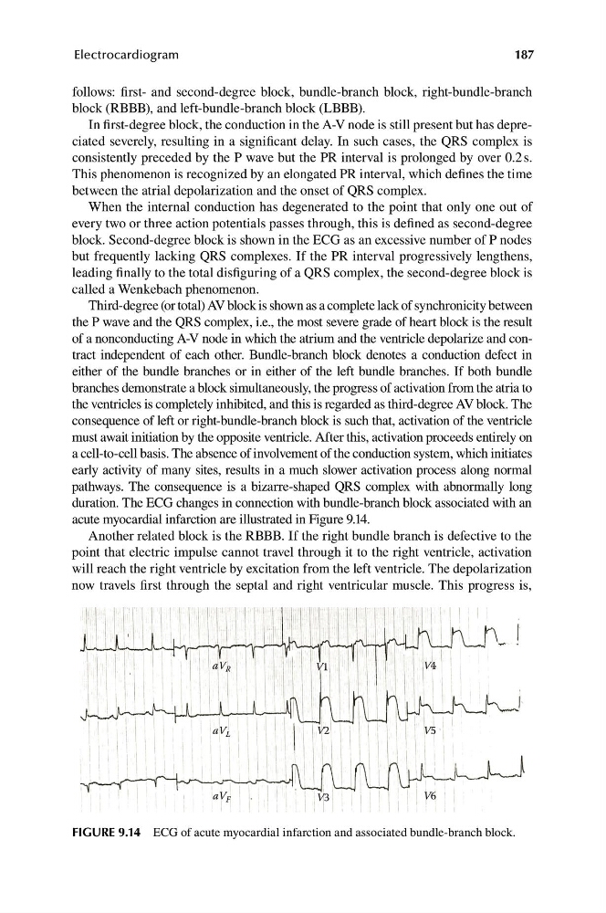
duration. The ECG changes in connection with bundle-branch block associated with an
acute myocardial infarction are illustrated in Figure 9.14.
Another related block is the RBBB. If the right bundle branch is defective to the
point that electric impulse cannot travel through it to the right ventricle, activation
will reach the right ventricle by excitation from the left ventricle. The depolarization
now travels first through the septal and right ventricular muscle. This progress is,
aV R
V1
V4
aV L
V2
V5
aV F
V3
V6
FIGURE 9.14 ECG of acute myocardial infarction and associated bundle-branch block.
Electrocardiogram
follows: first- and second-degree block, bundle-branch block, right-bundle-branch
block (RBBB), and left-bundle-branch block (LBBB).
In first-degree block, the conduction in the A-V node is still present but has depreciated severely, resulting in a significant delay. In such cases, the QRS complex is
consistently preceded by the P wave but the PR interval is prolonged by over 0.2 s.
This phenomenon is recognized by an elongated PR interval, which defines the time
between the atrial depolarization and the onset of QRS complex.
When the internal conduction has degenerated to the point that only one out of
every two or three action potentials passes through, this is defined as second-degree
block. Second-degree block is shown in the ECG as an excessive number of P nodes
but frequently lacking QRS complexes. If the PR interval progressively lengthens,
leading finally to the total disfiguring of a QRS complex, the second-degree block is
called a Wenkebach phenomenon.
Third-degree (or total) AV block is shown as a complete lack of synchronicity between
the P wave and the QRS complex, i.e., the most severe grade of heart block is the result
of a nonconducting A-V node in which the atrium and the ventricle depolarize and contract independent of each other. Bundle-branch block denotes a conduction defect in
either of the bundle branches or in either of the left bundle branches. If both bundle
branches demonstrate a block simultaneously, the progress of activation from the atria to
the ventricles is completely inhibited, and this is regarded as third-degree AV block. The
consequence of left or right-bundle-branch block is such that, activation of the ventricle
must await initiation by the opposite ventricle. After this, activation proceeds entirely on
a cell-to-cell basis. The absence of involvement of the conduction system, which initiates
early activity of many sites, results in a much slower activation process along normal
pathways. The consequence is a bizarre-shaped QRS complex with abnormally long
duration. The ECG changes in connection with bundle-branch block associated with an
acute myocardial infarction are illustrated in Figure 9.14.
Another related block is the RBBB. If the right bundle branch is defective to the
point that electric impulse cannot travel through it to the right ventricle, activation
will reach the right ventricle by excitation from the left ventricle. The depolarization
now travels first through the septal and right ventricular muscle. This progress is,
aV R
V1
V4
aV L
V2
V5
aV F
V3
V6
FIGURE 9.14 ECG of acute myocardial infarction and associated bundle-branch block.