

186
Biomedical Signal and Image Processing
Another frequent form of reentry is a short circuit in the Purkinje fibers at the end
of His bundle. The signal traveling through the His bundle does not conduct through
one of the branches in one direction but will allow conduction in the opposite direction, providing a secondary activation of the ventricles through the healthy His
branch. Cardiac reentry is one of the main causes of ventricular arrhythmias.
9.4.8 ATRIOVENTRICULAR BLOCK
As mentioned earlier, the A-V node is slower than the S-A node, and, as a result of
various illnesses, the conduction from S-A node to A-V node can be interrupted.
This is called AV block. Under these circumstances, the atria will contract faster
than the ventricles and the pump function will be severely compromised. As discussed earlier, if the P wave precedes the QRS complex with a PR interval of 0.12 s,
the AV conduction is normal. If the PR interval is fixed but shorter than normal,
either the origin of the impulse is closer to the ventricles or the AV conduction is
utilizing an (abnormal) bypass tract leading to preexcitation of the ventricles. The
latter is called the Wolff–Parkinson–White (WPW) syndrome and is discussed later.
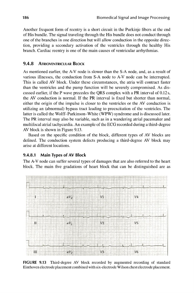
The PR interval may also be variable, such as in a wandering atrial pacemaker and
multifocal atrial tachycardia. An example of the ECG recorded during a third-degree
AV block is shown in Figure 9.13.
Based on the specific condition of the block, different types of AV blocks are
defined. The conduction system defects producing a third-degree AV block may
arise at different locations.
9.4.8.1 Main Types of AV Block
The A-V node can suffer several types of damages that are also referred to the heart
block. The main five gradations of heart block that can be distinguished are as
I
aV R
V1
V4
II
aV L
V2
V5
III
aV F
V3
V6
FIGURE 9.13 Third-degree AV block recorded by augmented recording of standard
Einthoven electrode placement combined with six-electrode Wilson chest electrode placement.
Biomedical Signal and Image Processing
Another frequent form of reentry is a short circuit in the Purkinje fibers at the end
of His bundle. The signal traveling through the His bundle does not conduct through
one of the branches in one direction but will allow conduction in the opposite direction, providing a secondary activation of the ventricles through the healthy His
branch. Cardiac reentry is one of the main causes of ventricular arrhythmias.
9.4.8 ATRIOVENTRICULAR BLOCK
As mentioned earlier, the A-V node is slower than the S-A node, and, as a result of
various illnesses, the conduction from S-A node to A-V node can be interrupted.
This is called AV block. Under these circumstances, the atria will contract faster
than the ventricles and the pump function will be severely compromised. As discussed earlier, if the P wave precedes the QRS complex with a PR interval of 0.12 s,
the AV conduction is normal. If the PR interval is fixed but shorter than normal,
either the origin of the impulse is closer to the ventricles or the AV conduction is
utilizing an (abnormal) bypass tract leading to preexcitation of the ventricles. The
latter is called the Wolff–Parkinson–White (WPW) syndrome and is discussed later.
The PR interval may also be variable, such as in a wandering atrial pacemaker and
multifocal atrial tachycardia. An example of the ECG recorded during a third-degree
AV block is shown in Figure 9.13.
Based on the specific condition of the block, different types of AV blocks are
defined. The conduction system defects producing a third-degree AV block may
arise at different locations.
9.4.8.1 Main Types of AV Block
The A-V node can suffer several types of damages that are also referred to the heart
block. The main five gradations of heart block that can be distinguished are as
I
aV R
V1
V4
II
aV L
V2
V5
III
aV F
V3
V6
FIGURE 9.13 Third-degree AV block recorded by augmented recording of standard
Einthoven electrode placement combined with six-electrode Wilson chest electrode placement.