

179
Electrocardiogram
V1
V2
V3
V4 V5
V6
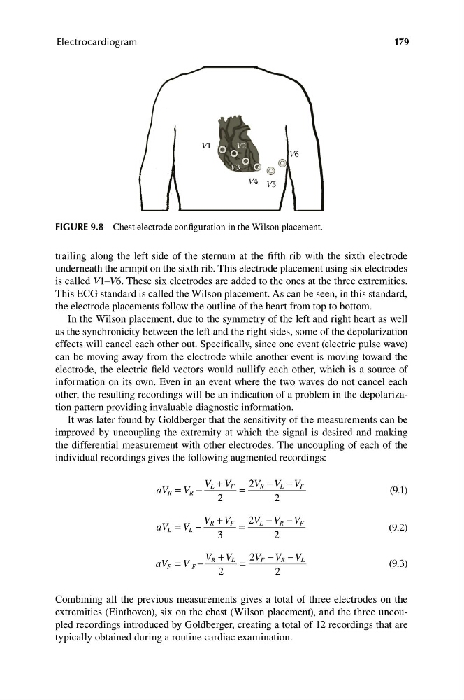
FIGURE 9.8 Chest electrode configuration in the Wilson placement.
trailing along the left side of the sternum at the fifth rib with the sixth electrode
underneath the armpit on the sixth rib. This electrode placement using six electrodes
is called V1–V6. These six electrodes are added to the ones at the three extremities.
This ECG standard is called the Wilson placement. As can be seen, in this standard,
the electrode placements follow the outline of the heart from top to bottom.
In the Wilson placement, due to the symmetry of the left and right heart as well
as the synchronicity between the left and the right sides, some of the depolarization
effects will cancel each other out. Specifically, since one event (electric pulse wave)
can be moving away from the electrode while another event is moving toward the
electrode, the electric field vectors would nullify each other, which is a source of
information on its own. Even in an event where the two waves do not cancel each
other, the resulting recordings will be an indication of a problem in the depolarization pattern providing invaluable diagnostic information.
It was later found by Goldberger that the sensitivity of the measurements can be
improved by uncoupling the extremity at which the signal is desired and making
the differential measurement with other electrodes. The uncoupling of each of the
individual recordings gives the following augmented recordings:
V L + V F 2V R − V L − V F
aV R = V R −
=
(9.1)
2
2
V R + V F 2V L − V R − V F
aV L = V L −
=
(9.2)
3
2
V R + V L 2V F − V R − V L
aV F = V F −
=
(9.3)
2
2
Combining all the previous measurements gives a total of three electrodes on the
extremities (Einthoven), six on the chest (Wilson placement), and the three uncoupled recordings introduced by Goldberger, creating a total of 12 recordings that are
typically obtained during a routine cardiac examination.
Electrocardiogram
V1
V2
V3
V4 V5
V6
FIGURE 9.8 Chest electrode configuration in the Wilson placement.
trailing along the left side of the sternum at the fifth rib with the sixth electrode
underneath the armpit on the sixth rib. This electrode placement using six electrodes
is called V1–V6. These six electrodes are added to the ones at the three extremities.
This ECG standard is called the Wilson placement. As can be seen, in this standard,
the electrode placements follow the outline of the heart from top to bottom.
In the Wilson placement, due to the symmetry of the left and right heart as well
as the synchronicity between the left and the right sides, some of the depolarization
effects will cancel each other out. Specifically, since one event (electric pulse wave)
can be moving away from the electrode while another event is moving toward the
electrode, the electric field vectors would nullify each other, which is a source of
information on its own. Even in an event where the two waves do not cancel each
other, the resulting recordings will be an indication of a problem in the depolarization pattern providing invaluable diagnostic information.
It was later found by Goldberger that the sensitivity of the measurements can be
improved by uncoupling the extremity at which the signal is desired and making
the differential measurement with other electrodes. The uncoupling of each of the
individual recordings gives the following augmented recordings:
V L + V F 2V R − V L − V F
aV R = V R −
=
(9.1)
2
2
V R + V F 2V L − V R − V F
aV L = V L −
=
(9.2)
3
2
V R + V L 2V F − V R − V L
aV F = V F −
=
(9.3)
2
2
Combining all the previous measurements gives a total of three electrodes on the
extremities (Einthoven), six on the chest (Wilson placement), and the three uncoupled recordings introduced by Goldberger, creating a total of 12 recordings that are
typically obtained during a routine cardiac examination.