

V L
V F
II
I
III
V R
178
Biomedical Signal and Image Processing
fact that the repolarization wave front is significantly longer in time duration than
the depolarization wave front. The repolarization also has a much smoother action
potential gradient, which incorporates a smaller gradient in the time derivative of the
cell membrane potential.
The ventricular repolarization is represented in the T wave of the ECG signal. The
T wave has a smaller amplitude than the QRS complex. If present, the U peak also
represents a portion of the ventricular repolarization.
Because of the electric nature of the heart muscle, the depolarization and the
repolarization are separated by a relatively long time period. As a result, in the electric recording of the heart activity, the expected triphasic action potential phenomenon will never be seen.
9.3.2 ECG ELECTRODE PLACEMENT
The ECG is not measured directly on the heart itself but on the exterior of the body.
Various internationally accepted electrode placements are in existence, which are
described next.
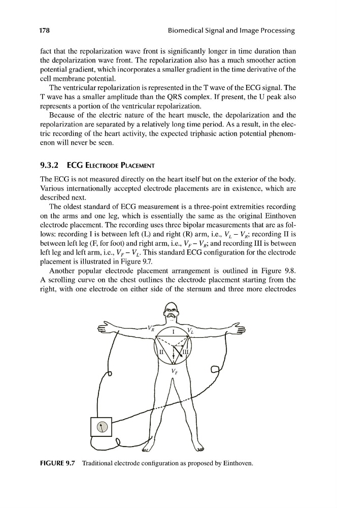
The oldest standard of ECG measurement is a three-point extremities recording
on the arms and one leg, which is essentially the same as the original Einthoven
electrode placement. The recording uses three bipolar measurements that are as follows: recording I is between left (L) and right (R) arm, i.e., V L − V R ; recording II is
between left leg (F, for foot) and right arm, i.e., V F − V R ; and recording III is between
left leg and left arm, i.e., V F − V L . This standard ECG configuration for the electrode
placement is illustrated in Figure 9.7.
Another popular electrode placement arrangement is outlined in Figure 9.8.
A scrolling curve on the chest outlines the electrode placement starting from the
right, with one electrode on either side of the sternum and three more electrodes
FIGURE 9.7 Traditional electrode configuration as proposed by Einthoven.
V F
II
I
III
V R
178
Biomedical Signal and Image Processing
fact that the repolarization wave front is significantly longer in time duration than
the depolarization wave front. The repolarization also has a much smoother action
potential gradient, which incorporates a smaller gradient in the time derivative of the
cell membrane potential.
The ventricular repolarization is represented in the T wave of the ECG signal. The
T wave has a smaller amplitude than the QRS complex. If present, the U peak also
represents a portion of the ventricular repolarization.
Because of the electric nature of the heart muscle, the depolarization and the
repolarization are separated by a relatively long time period. As a result, in the electric recording of the heart activity, the expected triphasic action potential phenomenon will never be seen.
9.3.2 ECG ELECTRODE PLACEMENT
The ECG is not measured directly on the heart itself but on the exterior of the body.
Various internationally accepted electrode placements are in existence, which are
described next.
The oldest standard of ECG measurement is a three-point extremities recording
on the arms and one leg, which is essentially the same as the original Einthoven
electrode placement. The recording uses three bipolar measurements that are as follows: recording I is between left (L) and right (R) arm, i.e., V L − V R ; recording II is
between left leg (F, for foot) and right arm, i.e., V F − V R ; and recording III is between
left leg and left arm, i.e., V F − V L . This standard ECG configuration for the electrode
placement is illustrated in Figure 9.7.
Another popular electrode placement arrangement is outlined in Figure 9.8.
A scrolling curve on the chest outlines the electrode placement starting from the
right, with one electrode on either side of the sternum and three more electrodes
FIGURE 9.7 Traditional electrode configuration as proposed by Einthoven.