

Intercalated disk
175
Electrocardiogram
producing impulses in a regular fashion to excite the atrium first, before trickling
down to the ventricle through the A-V node. The S-A node is tunable by hormonal
and parasympathetic nerve impulses.
The A-V node is positioned in the part of the septum close to the base of the
heart, i.e., the border between the atria and the ventricles, just above a section called
the annulus fibrosus. The annulus fibrosus is a connective tissue that separates the
atria from the ventricles and is an electric insulator. The A-V node is also under
the endocardium of the right atrium. This node not only generates impulses under
stimulus from the atrial depolarization but also has a depolarization sequence of
its own since it is constructed of nodal tissue as well. The depolarization rate of the
A-V node is generally slower than that of the S-A node. The impulse generated by
the A-V node is conducted through a bundle of nodal cells called the His bundle
that branch into multiple branches of conducting fibers that carry the depolarization impulse to the apex of the heart, the bottom of the heart, farthest away from
the outlet valves of the ventricle, which are at the base of the atrium again. The
branches of fibers spreading out from the His bundle are called Purkinje fibers.
The A-V node has a range of cells with different depolarization speeds. The cells
of the A-V node farthest from the His bundle are the fastest depolarizing, and the
lower ones are closer to the contact with the His bundle. The His bundle acts solely
as conductor of electric pulses. There are many terminal points of the Purkinje
fibers at the apex of the heart, ensuring a massive contractile motion at the tip. The polarization propagation speed is approximately 0.5 m/s for the cardiac muscle tissue, while
the propagation speed for the His bundle is approximately 2 m/s.
As mentioned earlier, the cardiac muscle is different from most other cells; in the
fact, that the cells themselves pass the depolarization signal on to only certain neighboring cells. All cardiac muscle cells in turn are connected to each other by a conducting
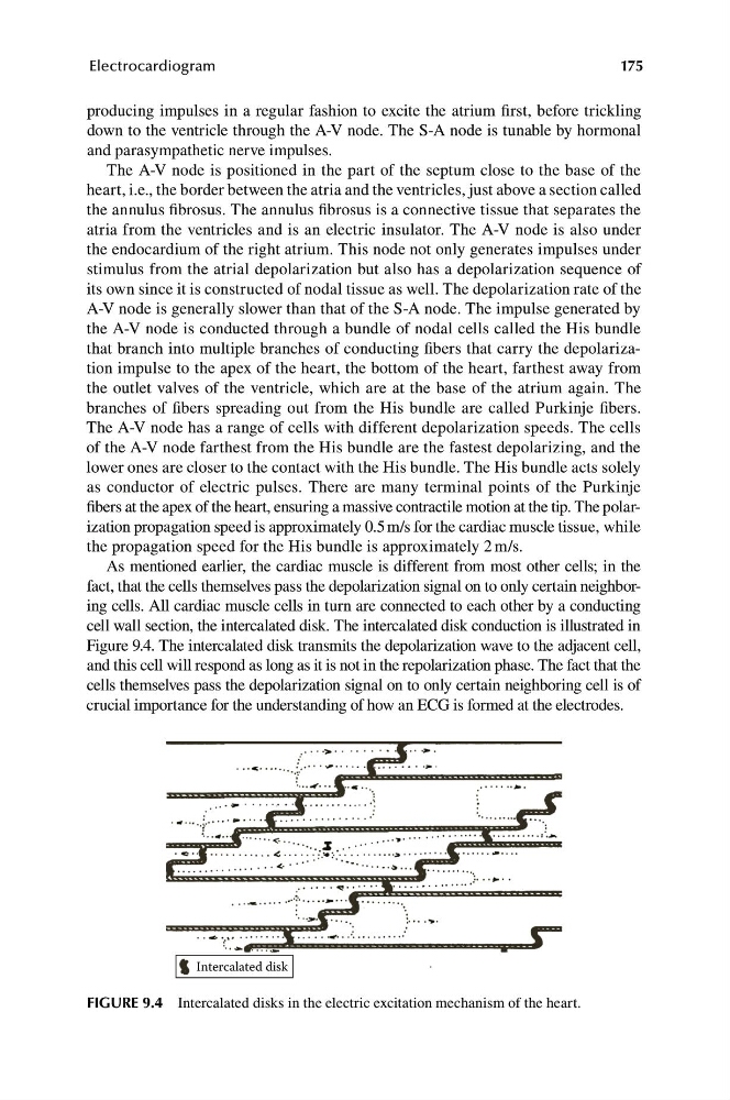
cell wall section, the intercalated disk. The intercalated disk conduction is illustrated in
Figure 9.4. The intercalated disk transmits the depolarization wave to the adjacent cell,
and this cell will respond as long as it is not in the repolarization phase. The fact that the
cells themselves pass the depolarization signal on to only certain neighboring cell is of
crucial importance for the understanding of how an ECG is formed at the electrodes.
FIGURE 9.4 Intercalated disks in the electric excitation mechanism of the heart.
175
Electrocardiogram
producing impulses in a regular fashion to excite the atrium first, before trickling
down to the ventricle through the A-V node. The S-A node is tunable by hormonal
and parasympathetic nerve impulses.
The A-V node is positioned in the part of the septum close to the base of the
heart, i.e., the border between the atria and the ventricles, just above a section called
the annulus fibrosus. The annulus fibrosus is a connective tissue that separates the
atria from the ventricles and is an electric insulator. The A-V node is also under
the endocardium of the right atrium. This node not only generates impulses under
stimulus from the atrial depolarization but also has a depolarization sequence of
its own since it is constructed of nodal tissue as well. The depolarization rate of the
A-V node is generally slower than that of the S-A node. The impulse generated by
the A-V node is conducted through a bundle of nodal cells called the His bundle
that branch into multiple branches of conducting fibers that carry the depolarization impulse to the apex of the heart, the bottom of the heart, farthest away from
the outlet valves of the ventricle, which are at the base of the atrium again. The
branches of fibers spreading out from the His bundle are called Purkinje fibers.
The A-V node has a range of cells with different depolarization speeds. The cells
of the A-V node farthest from the His bundle are the fastest depolarizing, and the
lower ones are closer to the contact with the His bundle. The His bundle acts solely
as conductor of electric pulses. There are many terminal points of the Purkinje
fibers at the apex of the heart, ensuring a massive contractile motion at the tip. The polarization propagation speed is approximately 0.5 m/s for the cardiac muscle tissue, while
the propagation speed for the His bundle is approximately 2 m/s.
As mentioned earlier, the cardiac muscle is different from most other cells; in the
fact, that the cells themselves pass the depolarization signal on to only certain neighboring cells. All cardiac muscle cells in turn are connected to each other by a conducting
cell wall section, the intercalated disk. The intercalated disk conduction is illustrated in
Figure 9.4. The intercalated disk transmits the depolarization wave to the adjacent cell,
and this cell will respond as long as it is not in the repolarization phase. The fact that the
cells themselves pass the depolarization signal on to only certain neighboring cell is of
crucial importance for the understanding of how an ECG is formed at the electrodes.
FIGURE 9.4 Intercalated disks in the electric excitation mechanism of the heart.