

174
AV-node
His
bundle
SA-node
Biomedical Signal and Image Processing
The remarkable difference with skeletal muscle in organizational structure is the
presence of a wire mesh of excitable cells: nodal tissue, running through the cardiac
muscle tissue. This network of excitable cells is in charge of control and adjustment
of the heartbeat and is further described in the next section.
9.2.2 CARDIAC EXCITATION PROCESS
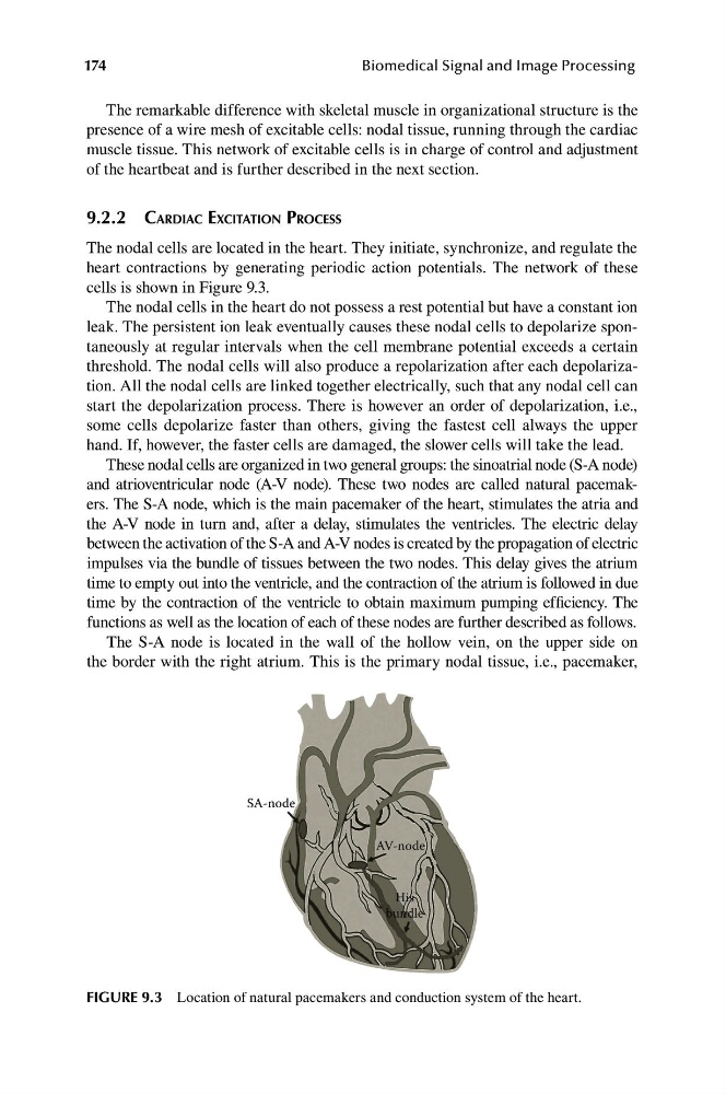
The nodal cells are located in the heart. They initiate, synchronize, and regulate the
heart contractions by generating periodic action potentials. The network of these
cells is shown in Figure 9.3.
The nodal cells in the heart do not possess a rest potential but have a constant ion
leak. The persistent ion leak eventually causes these nodal cells to depolarize spontaneously at regular intervals when the cell membrane potential exceeds a certain
threshold. The nodal cells will also produce a repolarization after each depolarization. All the nodal cells are linked together electrically, such that any nodal cell can
start the depolarization process. There is however an order of depolarization, i.e.,
some cells depolarize faster than others, giving the fastest cell always the upper
hand. If, however, the faster cells are damaged, the slower cells will take the lead.
These nodal cells are organized in two general groups: the sinoatrial node (S-A node)
and atrioventricular node (A-V node). These two nodes are called natural pacemakers. The S-A node, which is the main pacemaker of the heart, stimulates the atria and
the A-V node in turn and, after a delay, stimulates the ventricles. The electric delay
between the activation of the S-A and A-V nodes is created by the propagation of electric
impulses via the bundle of tissues between the two nodes. This delay gives the atrium
time to empty out into the ventricle, and the contraction of the atrium is followed in due
time by the contraction of the ventricle to obtain maximum pumping efficiency. The
functions as well as the location of each of these nodes are further described as follows.
The S-A node is located in the wall of the hollow vein, on the upper side on
the border with the right atrium. This is the primary nodal tissue, i.e., pacemaker,
FIGURE 9.3 Location of natural pacemakers and conduction system of the heart.
AV-node
His
bundle
SA-node
Biomedical Signal and Image Processing
The remarkable difference with skeletal muscle in organizational structure is the
presence of a wire mesh of excitable cells: nodal tissue, running through the cardiac
muscle tissue. This network of excitable cells is in charge of control and adjustment
of the heartbeat and is further described in the next section.
9.2.2 CARDIAC EXCITATION PROCESS
The nodal cells are located in the heart. They initiate, synchronize, and regulate the
heart contractions by generating periodic action potentials. The network of these
cells is shown in Figure 9.3.
The nodal cells in the heart do not possess a rest potential but have a constant ion
leak. The persistent ion leak eventually causes these nodal cells to depolarize spontaneously at regular intervals when the cell membrane potential exceeds a certain
threshold. The nodal cells will also produce a repolarization after each depolarization. All the nodal cells are linked together electrically, such that any nodal cell can
start the depolarization process. There is however an order of depolarization, i.e.,
some cells depolarize faster than others, giving the fastest cell always the upper
hand. If, however, the faster cells are damaged, the slower cells will take the lead.
These nodal cells are organized in two general groups: the sinoatrial node (S-A node)
and atrioventricular node (A-V node). These two nodes are called natural pacemakers. The S-A node, which is the main pacemaker of the heart, stimulates the atria and
the A-V node in turn and, after a delay, stimulates the ventricles. The electric delay
between the activation of the S-A and A-V nodes is created by the propagation of electric
impulses via the bundle of tissues between the two nodes. This delay gives the atrium
time to empty out into the ventricle, and the contraction of the atrium is followed in due
time by the contraction of the ventricle to obtain maximum pumping efficiency. The
functions as well as the location of each of these nodes are further described as follows.
The S-A node is located in the wall of the hollow vein, on the upper side on
the border with the right atrium. This is the primary nodal tissue, i.e., pacemaker,
FIGURE 9.3 Location of natural pacemakers and conduction system of the heart.