

96
Biomedical Signal and Image Processing
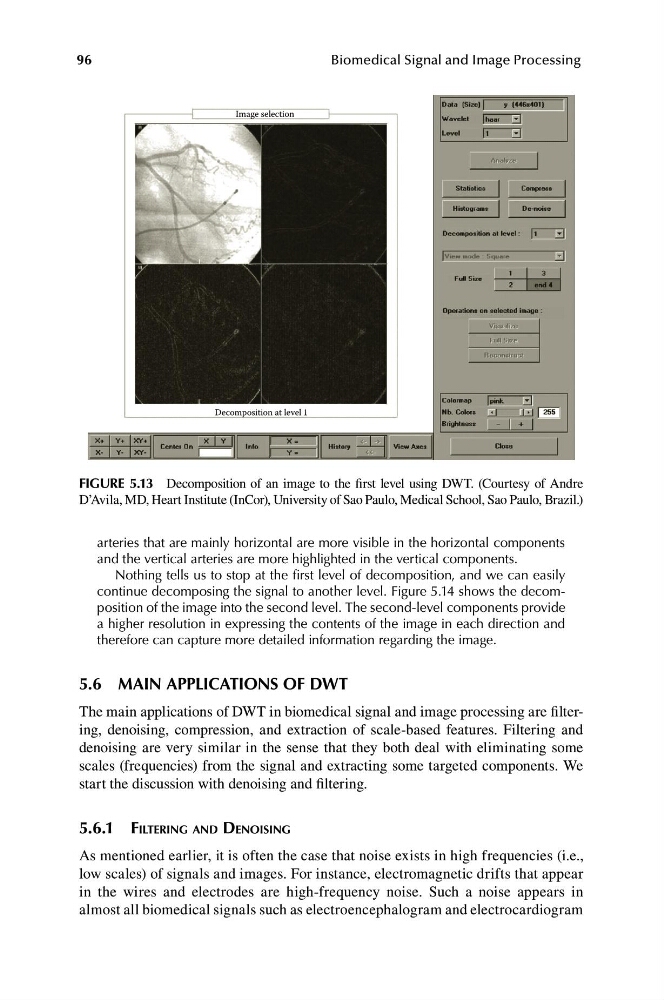
Image selection
Decomposition at level 1
FIGURE 5.13 Decomposition of an image to the first level using DWT. (Courtesy of Andre
D’Avila, MD, Heart Institute (InCor), University of Sao Paulo, Medical School, Sao Paulo, Brazil.)
arteries that are mainly horizontal are more visible in the horizontal components
and the vertical arteries are more highlighted in the vertical components.
Nothing tells us to stop at the first level of decomposition, and we can easily
continue decomposing the signal to another level. Figure 5.14 shows the decomposition of the image into the second level. The second-level components provide
a higher resolution in expressing the contents of the image in each direction and
therefore can capture more detailed information regarding the image.
5.6 MAIN APPLICATIONS OF DWT
The main applications of DWT in biomedical signal and image processing are filtering, denoising, compression, and extraction of scale-based features. Filtering and
denoising are very similar in the sense that they both deal with eliminating some
scales (frequencies) from the signal and extracting some targeted components. We
start the discussion with denoising and filtering.
5.6.1 FILTERING AND DENOISING
As mentioned earlier, it is often the case that noise exists in high frequencies (i.e.,
low scales) of signals and images. For instance, electromagnetic drifts that appear
in the wires and electrodes are high-frequency noise. Such a noise appears in
almost all biomedical signals such as electroencephalogram and electrocardiogram
Biomedical Signal and Image Processing
Image selection
Decomposition at level 1
FIGURE 5.13 Decomposition of an image to the first level using DWT. (Courtesy of Andre
D’Avila, MD, Heart Institute (InCor), University of Sao Paulo, Medical School, Sao Paulo, Brazil.)
arteries that are mainly horizontal are more visible in the horizontal components
and the vertical arteries are more highlighted in the vertical components.
Nothing tells us to stop at the first level of decomposition, and we can easily
continue decomposing the signal to another level. Figure 5.14 shows the decomposition of the image into the second level. The second-level components provide
a higher resolution in expressing the contents of the image in each direction and
therefore can capture more detailed information regarding the image.
5.6 MAIN APPLICATIONS OF DWT
The main applications of DWT in biomedical signal and image processing are filtering, denoising, compression, and extraction of scale-based features. Filtering and
denoising are very similar in the sense that they both deal with eliminating some
scales (frequencies) from the signal and extracting some targeted components. We
start the discussion with denoising and filtering.
5.6.1 FILTERING AND DENOISING
As mentioned earlier, it is often the case that noise exists in high frequencies (i.e.,
low scales) of signals and images. For instance, electromagnetic drifts that appear
in the wires and electrodes are high-frequency noise. Such a noise appears in
almost all biomedical signals such as electroencephalogram and electrocardiogram