

42
2 Dopachrome Conversion
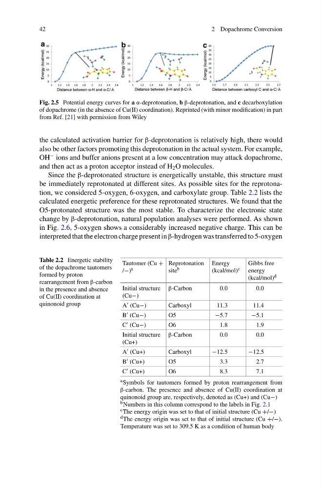
Fig. 2.5 Potential energy curves for a α-deprotonation, b β-deprotonation, and c decarboxylation
of dopachrome (in the absence of Cu(II) coordination). Reprinted (with minor modification) in part
from Ref. [21] with permission from Wiley
the calculated activation barrier for β-deprotonation is relatively high, there would
also be other factors promoting this deprotonation in the actual system. For example,
OH
− ions and buffer anions present at a low concentration may attack dopachrome,
and then act as a proton acceptor instead of H 2 O molecules.
Since the β-deprotonated structure is energetically unstable, this structure must
be immediately reprotonated at different sites. As possible sites for the reprotonation, we considered 5-oxygen, 6-oxygen, and carboxylate group. Table 2.2 lists the
calculated energetic preference for these reprotonated structures. We found that the
O5-protonated structure was the most stable. To characterize the electronic state
change by β-deprotonation, natural population analyses were performed. As shown
in Fig. 2.6, 5-oxygen shows a considerably increased negative charge. This can be
interpreted that the electron charge present in β-hydrogen was transferred to 5-oxygen
Table 2.2 Energetic stability
of the dopachrome tautomers
formed by proton
rearrangement from β-carbon
in the presence and absence
of Cu(II) coordination at
quinonoid group
Tautomer (Cu +
/−) a
Reprotonation
site b
Energy
(kcal/mol) c
Gibbs free
energy
(kcal/mol) d
Initial structure
(Cu−)
β-Carbon
0.0
0.0
A (Cu−)
Carboxyl
11.3
11.4
B (Cu−)
O5
−5.7
−5.1
C (Cu−)
O6
1.8
1.9
Initial structure
(Cu+)
β-Carbon
0.0
0.0
A (Cu+)
Carboxyl
−12.5
−12.5
B (Cu+)
O5
3.3
2.7
C (Cu+)
O6
8.3
7.1
a Symbols for tautomers formed by proton rearrangement from
β-carbon. The presence and absence of Cu(II) coordination at
quinonoid group are, respectively, denoted as (Cu+) and (Cu−)
b Numbers in this column correspond to the labels in Fig. 2.1
c The energy origin was set to that of initial structure (Cu +/−)
d The energy origin was set to that of initial structure (Cu +/−).
Temperature was set to 309.5 K as a condition of human body
2 Dopachrome Conversion
Fig. 2.5 Potential energy curves for a α-deprotonation, b β-deprotonation, and c decarboxylation
of dopachrome (in the absence of Cu(II) coordination). Reprinted (with minor modification) in part
from Ref. [21] with permission from Wiley
the calculated activation barrier for β-deprotonation is relatively high, there would
also be other factors promoting this deprotonation in the actual system. For example,
OH
− ions and buffer anions present at a low concentration may attack dopachrome,
and then act as a proton acceptor instead of H 2 O molecules.
Since the β-deprotonated structure is energetically unstable, this structure must
be immediately reprotonated at different sites. As possible sites for the reprotonation, we considered 5-oxygen, 6-oxygen, and carboxylate group. Table 2.2 lists the
calculated energetic preference for these reprotonated structures. We found that the
O5-protonated structure was the most stable. To characterize the electronic state
change by β-deprotonation, natural population analyses were performed. As shown
in Fig. 2.6, 5-oxygen shows a considerably increased negative charge. This can be
interpreted that the electron charge present in β-hydrogen was transferred to 5-oxygen
Table 2.2 Energetic stability
of the dopachrome tautomers
formed by proton
rearrangement from β-carbon
in the presence and absence
of Cu(II) coordination at
quinonoid group
Tautomer (Cu +
/−) a
Reprotonation
site b
Energy
(kcal/mol) c
Gibbs free
energy
(kcal/mol) d
Initial structure
(Cu−)
β-Carbon
0.0
0.0
A (Cu−)
Carboxyl
11.3
11.4
B (Cu−)
O5
−5.7
−5.1
C (Cu−)
O6
1.8
1.9
Initial structure
(Cu+)
β-Carbon
0.0
0.0
A (Cu+)
Carboxyl
−12.5
−12.5
B (Cu+)
O5
3.3
2.7
C (Cu+)
O6
8.3
7.1
a Symbols for tautomers formed by proton rearrangement from
β-carbon. The presence and absence of Cu(II) coordination at
quinonoid group are, respectively, denoted as (Cu+) and (Cu−)
b Numbers in this column correspond to the labels in Fig. 2.1
c The energy origin was set to that of initial structure (Cu +/−)
d The energy origin was set to that of initial structure (Cu +/−).
Temperature was set to 309.5 K as a condition of human body