

6
1 Melanin Chemistry
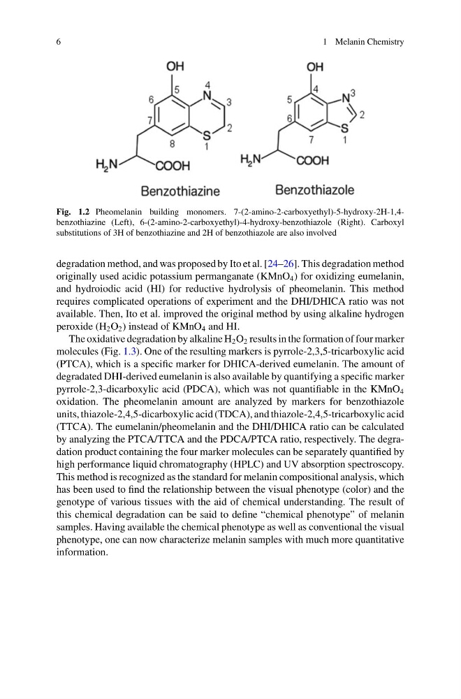
Fig. 1.2 Pheomelanin building monomers. 7-(2-amino-2-carboxyethyl)-5-hydroxy-2H-1,4benzothiazine (Left), 6-(2-amino-2-carboxyethyl)-4-hydroxy-benzothiazole (Right). Carboxyl
substitutions of 3H of benzothiazine and 2H of benzothiazole are also involved
degradation method, and was proposed by Ito et al. [24–26]. This degradation method
originally used acidic potassium permanganate (KMnO 4 ) for oxidizing eumelanin,
and hydroiodic acid (HI) for reductive hydrolysis of pheomelanin. This method
requires complicated operations of experiment and the DHI/DHICA ratio was not
available. Then, Ito et al. improved the original method by using alkaline hydrogen
peroxide (H 2 O 2 ) instead of KMnO 4 and HI.
The oxidative degradation by alkaline H 2 O 2 results in the formation of four marker
molecules (Fig. 1.3). One of the resulting markers is pyrrole-2,3,5-tricarboxylic acid
(PTCA), which is a specific marker for DHICA-derived eumelanin. The amount of
degradated DHI-derived eumelanin is also available by quantifying a specific marker
pyrrole-2,3-dicarboxylic acid (PDCA), which was not quantifiable in the KMnO 4
oxidation. The pheomelanin amount are analyzed by markers for benzothiazole
units, thiazole-2,4,5-dicarboxylic acid (TDCA), and thiazole-2,4,5-tricarboxylic acid
(TTCA). The eumelanin/pheomelanin and the DHI/DHICA ratio can be calculated
by analyzing the PTCA/TTCA and the PDCA/PTCA ratio, respectively. The degradation product containing the four marker molecules can be separately quantified by
high performance liquid chromatography (HPLC) and UV absorption spectroscopy.
This method is recognized as the standard for melanin compositional analysis, which
has been used to find the relationship between the visual phenotype (color) and the
genotype of various tissues with the aid of chemical understanding. The result of
this chemical degradation can be said to define “chemical phenotype” of melanin
samples. Having available the chemical phenotype as well as conventional the visual
phenotype, one can now characterize melanin samples with much more quantitative
information.
1 Melanin Chemistry
Fig. 1.2 Pheomelanin building monomers. 7-(2-amino-2-carboxyethyl)-5-hydroxy-2H-1,4benzothiazine (Left), 6-(2-amino-2-carboxyethyl)-4-hydroxy-benzothiazole (Right). Carboxyl
substitutions of 3H of benzothiazine and 2H of benzothiazole are also involved
degradation method, and was proposed by Ito et al. [24–26]. This degradation method
originally used acidic potassium permanganate (KMnO 4 ) for oxidizing eumelanin,
and hydroiodic acid (HI) for reductive hydrolysis of pheomelanin. This method
requires complicated operations of experiment and the DHI/DHICA ratio was not
available. Then, Ito et al. improved the original method by using alkaline hydrogen
peroxide (H 2 O 2 ) instead of KMnO 4 and HI.
The oxidative degradation by alkaline H 2 O 2 results in the formation of four marker
molecules (Fig. 1.3). One of the resulting markers is pyrrole-2,3,5-tricarboxylic acid
(PTCA), which is a specific marker for DHICA-derived eumelanin. The amount of
degradated DHI-derived eumelanin is also available by quantifying a specific marker
pyrrole-2,3-dicarboxylic acid (PDCA), which was not quantifiable in the KMnO 4
oxidation. The pheomelanin amount are analyzed by markers for benzothiazole
units, thiazole-2,4,5-dicarboxylic acid (TDCA), and thiazole-2,4,5-tricarboxylic acid
(TTCA). The eumelanin/pheomelanin and the DHI/DHICA ratio can be calculated
by analyzing the PTCA/TTCA and the PDCA/PTCA ratio, respectively. The degradation product containing the four marker molecules can be separately quantified by
high performance liquid chromatography (HPLC) and UV absorption spectroscopy.
This method is recognized as the standard for melanin compositional analysis, which
has been used to find the relationship between the visual phenotype (color) and the
genotype of various tissues with the aid of chemical understanding. The result of
this chemical degradation can be said to define “chemical phenotype” of melanin
samples. Having available the chemical phenotype as well as conventional the visual
phenotype, one can now characterize melanin samples with much more quantitative
information.